The FAT10/UBD Antibody (CAB5491) is a high-quality antibody developed for reliable detection and analysis of target proteins. This antibody, produced in rabbits, exhibits high specificity and sensitivity towards human samples, making it an ideal choice for Western blot applications. By binding to ubiquitin D, this antibody enables accurate detection and analysis in a variety of cell types, facilitating in-depth investigations into ubiquitin signaling pathways and their roles in cellular physiology.Ubiquitin D is a crucial player in the ubiquitin system, which regulates protein degradation, DNA repair, and various signaling pathways within the cell. Dysregulation of ubiquitin-mediated processes has been implicated in a wide range of diseases, including cancer, neurodegenerative disorders, and autoimmune diseases.
This antibody is validated for use in WB, IF/ICC, ELISA applications and has demonstrated reactivity against Human, Mouse samples.
Product Name:
FAT10/UBD Antibody
SKU:
CAB5491
Size:
20μL, 100μL
Reactivity:
Human, Mouse
Conjugate:
Unconjugated
Immunogen:
Recombinant protein (or fragment).This information is considered to be commercially sensitive.
Recommended starting concentration is 1 μg/mL. Please optimize the concentration based on your specific assay requirements.
Synonyms:
FAT10, UBD-3, GABBR1, FAT10/UBD
Positive Sample:
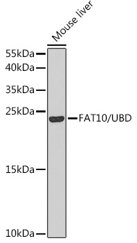
Mouse liver
Cellular Localization:
Cytoplasm, Nucleus.
Calculated MW:
18kDa
Observed MW:
24kDa
This gene encodes a protein which contains two ubiquitin-like domains and appears to have similar function to ubiquitin. Through covalent attachment, the encoded protein targets other proteins for 26S proteasome degradation. This protein has been implicated to function in many cellular processes, including caspase-dependent apoptosis, formation of aggresomes, mitotic regulation, and dendritic cell maturation. Upregulation of this gene may promote inflammation in chronic kidney disease and has been observed in many cancer types.
Purification Method
Affinity purification
Gene ID
10537
RRID
AB_2766290
Buffer Information
Store at -20℃. Avoid freeze / thaw cycles. Buffer: PBS containing 50% glycerol, preserved with proclin300 or sodium azide, pH 7.3.
Western blot analysis of lysates from mouse liver, using FAT10/UBD Rabbit pAb (CAB5491) at 1:1000 dilution. Secondary antibody: HRP-conjugated Goat anti-Rabbit IgG (H+L) (CABS014) at 1:10000 dilution. Lysates/proteins: 25μg per lane. Blocking buffer: 3% nonfat dry milk in TBST. Detection: ECL Basic Kit (AbGn00020). Exposure time: 60s.
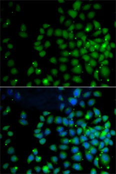
Immunofluorescence analysis of A-549 cells using FAT10/UBD Rabbit pAb (CAB5491). Secondary antibody: Cy3-conjugated Goat anti-Rabbit IgG (H+L) (CABS007) at 1:500 dilution. Blue: DAPI for nuclear staining.