The TSLP Antibody (CAB13134) is a high-quality antibody developed for reliable detection and analysis of target proteins. This antibody, raised in rabbits, is highly specific to human samples and has been validated for use in Western blot applications.TSLP is known for its ability to promote allergic inflammation and drive immune responses, making it a promising target for studies in allergy, asthma, and autoimmune diseases.
This antibody is validated for use in WB, IF/ICC, ELISA applications and has demonstrated reactivity against Human, Mouse, Rat samples.
Product Name:
TSLP Antibody
SKU:
CAB13134
Size:
20μL, 100μL
Reactivity:
Human, Mouse, Rat
Conjugate:
Unconjugated
Immunogen:
Recombinant protein (or fragment).This information is considered to be commercially sensitive.
Recommended starting concentration is 1 μg/mL. Please optimize the concentration based on your specific assay requirements.
Synonyms:
TSLP
Positive Sample:
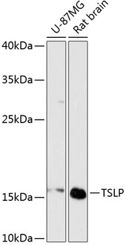
U-87MG, Rat brain
Cellular Localization:
Secreted.
Calculated MW:
18kDa
Observed MW:
18kDa
This gene encodes a hemopoietic cytokine proposed to signal through a heterodimeric receptor complex composed of the thymic stromal lymphopoietin receptor and the IL-7R alpha chain. It mainly impacts myeloid cells and induces the release of T cell-attracting chemokines from monocytes and enhances the maturation of CD11c(+) dendritic cells. The protein promotes T helper type 2 (TH2) cell responses that are associated with immunity in various inflammatory diseases, including asthma, allergic inflammation and chronic obstructive pulmonary disease. The protein is therefore considered a potential therapeutic target for the treatment of such diseases. In addition, the shorter (predominant) isoform is an antimicrobial protein, displaying antibacterial and antifungal activity against B. cereus, E. coli, E. faecalis, S. mitis, S. epidermidis, and C. albicans. Alternative splicing of this gene results in multiple transcript variants.
Purification Method
Affinity purification
Gene ID
85480
RRID
AB_2759984
Buffer Information
Store at -20℃. Avoid freeze / thaw cycles. Buffer: PBS containing 50% glycerol, preserved with proclin300 or sodium azide, pH 7.3.
Western blot analysis of various lysates using TSLP Rabbit pAb (CAB13134) at 1:3000 dilution. Secondary antibody: HRP-conjugated Goat anti-Rabbit IgG (H+L) (CABS014) at 1:10000 dilution. Lysates/proteins: 25μg per lane. Blocking buffer: 3% nonfat dry milk in TBST. Detection: ECL Basic Kit (AbGn00020). Exposure time: 90s.
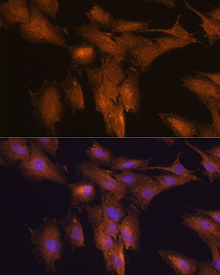
Immunofluorescence analysis of C6 cells using TSLP Rabbit pAb (CAB13134) at dilution of 1:100. Secondary antibody: Cy3-conjugated Goat anti-Rabbit IgG (H+L) (CABS007) at 1:500 dilution. Blue: DAPI for nuclear staining.
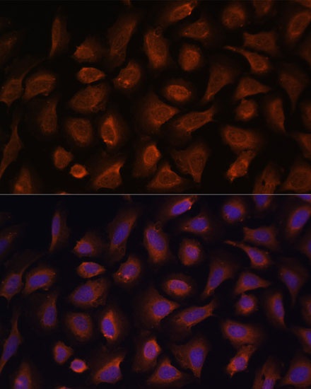
Immunofluorescence analysis of U-2 OS cells using TSLP Rabbit pAb (CAB13134) at dilution of 1:100. Secondary antibody: Cy3-conjugated Goat anti-Rabbit IgG (H+L) (CABS007) at 1:500 dilution. Blue: DAPI for nuclear staining.