The RB Antibody (CAB16966) is a high-quality antibody developed for reliable detection and analysis of target proteins. This antibody, produced in rabbits, exhibits high specificity and sensitivity when detecting RB1 in human samples, making it a reliable choice for Western blot applications.RB1 is a tumor suppressor protein that plays a crucial role in cell division and growth control. Mutations in the RB1 gene have been linked to various cancers, including retinoblastoma, osteosarcoma, and breast cancer. By targeting RB1 with this polyclonal antibody, researchers can accurately monitor RB1 expression levels in different cell types, providing valuable insights into cell cycle regulation and cancer progression.
This antibody is validated for use in WB, IF/ICC, ELISA applications and has demonstrated reactivity against Human samples.
Product Name:
RB Antibody
SKU:
CAB16966
Size:
20μL, 100μL
Reactivity:
Human
Immunogen:
Recombinant protein (or fragment).This information is considered to be commercially sensitive.
Recommended starting concentration is 1 μg/mL. Please optimize the concentration based on your specific assay requirements.
Synonyms:
RB, pRb, OSRC, pp110, p105-Rb, PPP1R130, p110-RB1
Positive Sample:
Jurkat
Cellular Localization:
Nucleus.
Calculated MW:
106kDa
Observed MW:
110kDa
The protein encoded by this gene is a negative regulator of the cell cycle and was the first tumor suppressor gene found. The encoded protein also stabilizes constitutive heterochromatin to maintain the overall chromatin structure. The active, hypophosphorylated form of the protein binds transcription factor E2F1. Defects in this gene are a cause of childhood cancer retinoblastoma (RB), bladder cancer, and osteogenic sarcoma.
Purification Method
Affinity purification
Gene ID
5925
RRID
AB_2771950
Buffer Information
Store at -20℃. Avoid freeze / thaw cycles. Buffer: PBS containing 50% glycerol, preserved with proclin300 or sodium azide, pH 7.3.
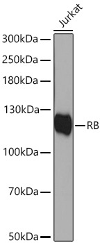
Western blot analysis of lysates from Jurkat cells using RB Rabbit pAb (CAB16966) at 1:1000 dilution incubated overnight at 4℃. Secondary antibody: HRP-conjugated Goat anti-Rabbit IgG (H+L) (CABS014) at 1:10000 dilution. Lysates/proteins: 25 μg per lane. Blocking buffer: 3% nonfat dry milk in TBST. Detection: ECL Basic Kit (AbGn00020). Exposure time: 90 s.
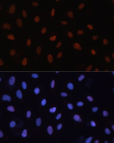
Immunofluorescence analysis of U-2 OS cells using RB Rabbit pAb (CAB16966) at dilution of 1:100. Secondary antibody: Cy3-conjugated Goat anti-Rabbit IgG (H+L) (CABS007) at 1:500 dilution. Blue: DAPI for nuclear staining.