The PTK7 Antibody (CAB9839) is a high-quality antibody developed for reliable detection and analysis of target proteins. This antibody, generated in rabbits, exhibits high reactivity with human samples and is validated for use in Western blot applications. By targeting the PTK7 protein, this antibody enables researchers to detect and analyze PTK7 expression in different cell types, making it well-suited for investigations in cancer biology and developmental biology.PTK7 plays a crucial role in cell signaling pathways and has been implicated in tumorigenesis, making it a promising target for cancer therapy research.
This antibody is validated for use in WB, IHC-P, ELISA applications and has demonstrated reactivity against Human, Mouse, Rat samples.
Product Name:
PTK7 Antibody
SKU:
CAB9839
Size:
20μL, 100μL
Reactivity:
Human, Mouse, Rat
Conjugate:
Unconjugated
Immunogen:
Recombinant protein (or fragment).This information is considered to be commercially sensitive.
Recommended starting concentration is 1 μg/mL. Please optimize the concentration based on your specific assay requirements.
Synonyms:
CCK4, CCK-4, PTK7
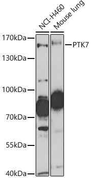
Positive Sample:
NCI-H460, Mouse lung
Cellular Localization:
Cell Junction, Membrane, Single-Pass Type I Membrane Protein.
Calculated MW:
118kDa
Observed MW:
160kDa
This gene encodes a member of the receptor protein tyrosine kinase family of proteins that transduce extracellular signals across the cell membrane. The encoded protein lacks detectable catalytic tyrosine kinase activity, is involved in the Wnt signaling pathway and plays a role in multiple cellular processes including polarity and adhesion. Alternatively spliced transcript variants encoding multiple isoforms have been observed for this gene.
Purification Method
Affinity purification
Gene ID
5754
RRID
AB_2771869
Buffer Information
Store at -20℃. Avoid freeze / thaw cycles. Buffer: PBS with 0.09% Sodium azide,50% glycerol,pH7.3.
Western blot analysis of various lysates using PTK7 Rabbit pAb (CAB9839) at 1:1000 dilution. Secondary antibody: HRP-conjugated Goat anti-Rabbit IgG (H+L) (CABS014) at 1:10000 dilution. Lysates/proteins: 25μg per lane. Blocking buffer: 3% nonfat dry milk in TBST. Detection: ECL Basic Kit (AbGn00020). Exposure time: 30s.
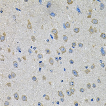
Immunohistochemistry analysis of paraffin-embedded Mouse brain using PTK7 Rabbit pAb (CAB9839) at dilution of 1:100 (40x lens). Microwave antigen retrieval performed with 0.01M PBS Buffer (pH 7.2) prior to IHC staining.
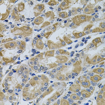
Immunohistochemistry analysis of paraffin-embedded Human stomach using PTK7 Rabbit pAb (CAB9839) at dilution of 1:100 (40x lens). Microwave antigen retrieval performed with 0.01M PBS Buffer (pH 7.2) prior to IHC staining.