The Phospho-MYL9-T18/S19 Antibody (CABP0955) is a high-quality antibody developed for reliable detection and analysis of target proteins. This antibody, generated in rabbits, is highly specific for detecting phosphorylation at threonine 18 and serine 19 residues of MYL9 in human samples. Validated for use in Western blot applications, it enables the detection and analysis of MYL9 phosphorylation levels in various cell types.MYL9, also known as myosin regulatory light chain 9, plays a crucial role in the control of actin-myosin interactions and cell contractility. Phosphorylation of MYL9 at threonine 18 and serine 19 sites is known to regulate its activity and impact cellular processes such as cell migration, adhesion, and cytokinesis.
This antibody is validated for use in WB, IHC-P, ELISA applications and has demonstrated reactivity against Human, Mouse, Rat samples.
Product Name:
Phospho-MYL9-T18/S19 Antibody
SKU:
CABP0955
Size:
20μL, 100μL
Reactivity:
Human, Mouse, Rat
Conjugate:
Unconjugated
Immunogen:
Synthetic peptide. This information is considered to be commercially sensitive.
Sequence:
RATS NV
Tested Applications:
WBIHC-PELISA
Recommended Dilution:
WB
1:1000 - 1:5000
IHC-P
1:50 - 1:200
ELISA
Recommended starting concentration is 1 μg/mL. Please optimize the concentration based on your specific assay requirements.
Cell Cortex, Cytoplasm, Cytosol, Myofibril, Z Disc.
Calculated MW:
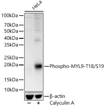
20kDa
Observed MW:
18kDa
Myosin, a structural component of muscle, consists of two heavy chains and four light chains. The protein encoded by this gene is a myosin light chain that may regulate muscle contraction by modulating the ATPase activity of myosin heads. The encoded protein binds calcium and is activated by myosin light chain kinase. Two transcript variants encoding different isoforms have been found for this gene.
Purification Method
Affinity purification
Gene ID
10398
RRID
AB_2863864
Buffer Information
Store at -20℃. Avoid freeze / thaw cycles. Buffer: PBS containing 50% glycerol, preserved with proclin300 or sodium azide, pH 7.3.
Western blot analysis of lysates from HeLa cells, using Phospho-MYL9-T18/S19 Rabbit pAb (CABP0955) at 1:3000 dilution. Secondary antibody: HRP-conjugated Goat anti-Rabbit IgG (H+L) (CABS014) at 1:10000 dilution. Lysates/proteins: 25μg per lane. Blocking buffer: 3% nonfat dry milk in TBST. Detection: ECL Basic Kit (AbGn00020). Exposure time: 10s.
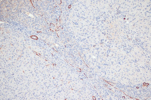
Immunohistochemistry analysis of paraffin-embedded Rat ovary using Phospho-MYL9-T18/S19 Rabbit pAb (CABP0955) at dilution of 1:100 (20x lens). Microwave antigen retrieval performed with 0.01M Tris/EDTA Buffer (pH 9.0) prior to IHC staining.