The Phospho-ERK1-Y204 Antibody (CABP0235) is a high-quality antibody developed for reliable detection and analysis of target proteins. This antibody, produced in rabbits, specifically targets the phosphorylated form of MAPK3 at tyrosine 204 and is highly reactive with human samples.Validated for use in Western blot applications, this antibody binds to the phosphorylated MAPK3 protein, allowing for precise detection and analysis in different cell types. Its utility in immunology and cancer research is particularly notable, as MAPK3 is known to play a significant role in regulating immune responses and tumor cell growth.
This antibody is validated for use in WB, IHC-P, IF/ICC, IP, ELISA applications and has demonstrated reactivity against Human, Mouse, Rat samples.
Product Name:
Phospho-ERK1-Y204 Antibody
SKU:
CABP0235
Size:
20μL, 100μL
Reactivity:
Human, Mouse, Rat
Conjugate:
Unconjugated
Immunogen:
Synthetic peptide. This information is considered to be commercially sensitive.
Sequence:
TEYV A
Tested Applications:
WBIHC-PIF/ICCIPELISA
Recommended Dilution:
WB
1:500 - 1:1000
IHC-P
1:50 - 1:100
IF/ICC
1:100 - 1:200
IP
0.5μg-4μg antibody for 200μg-400μg extracts of whole cells
ELISA
Recommended starting concentration is 1 μg/mL. Please optimize the concentration based on your specific assay requirements.
The protein encoded by this gene is a member of the MAP kinase family. MAP kinases, also known as extracellular signal-regulated kinases (ERKs), act in a signaling cascade that regulates various cellular processes such as proliferation, differentiation, and cell cycle progression in response to a variety of extracellular signals. This kinase is activated by upstream kinases, resulting in its translocation to the nucleus where it phosphorylates nuclear targets. Alternatively spliced transcript variants encoding different protein isoforms have been described.
Purification Method
Affinity purification
Gene ID
5595
RRID
AB_2771314
Buffer Information
Store at -20℃. Avoid freeze / thaw cycles. Buffer: PBS containing 50% glycerol, preserved with proclin300 or sodium azide, pH 7.3.
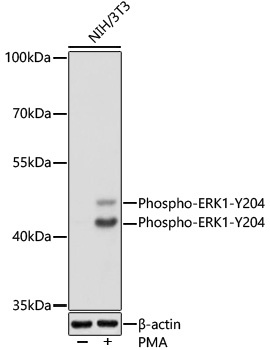
Western blot analysis of lysates from NIH/3T3 cells using Phospho-ERK1-Y204 Rabbit pAb (CABP0235) at 1:1000 dilution incubated overnight at 4℃. NIH/3T3 cells were treated with PMA/TPA (200 nM) at 37℃ for 30 minutes after serum-starvation overnight. Secondary antibody: HRP-conjugated Goat anti-Rabbit IgG (H+L) (CABS014) at 1:10000 dilution. Lysates/proteins: 25μg per lane. Blocking buffer: 3% nonfat dry milk in TBST. Detection: ECL Basic Kit (AbGn00020). Exposure time: 1s.
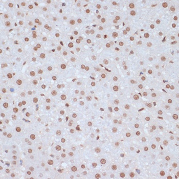
Immunohistochemistry analysis of paraffin-embedded Rat liver using Phospho-ERK1-Y204 Rabbit pAb (CABP0235) at dilution of 1:100 (40x lens). Microwave antigen retrieval performed with 0.01M Tris/EDTA Buffer (pH 9.0) prior to IHC staining.
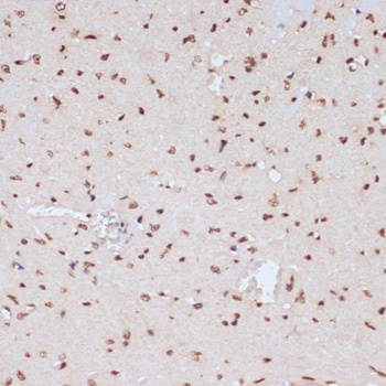
Immunohistochemistry analysis of paraffin-embedded Mouse heart using Phospho-ERK1-Y204 Rabbit pAb (CABP0235) at dilution of 1:100 (40x lens). Microwave antigen retrieval performed with 0.01M Tris/EDTA Buffer (pH 9.0) prior to IHC staining.
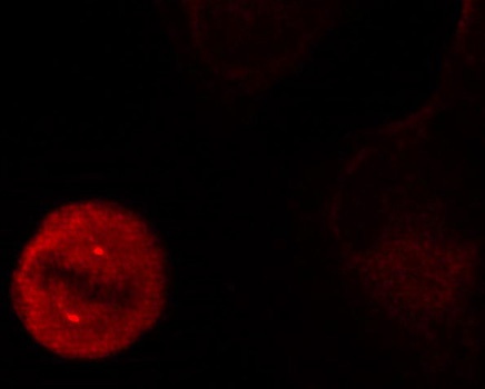
Immunofluorescence analysis of methanol-fixed HeLa cells showing centrosome and nuclear staining using Phospho-ERK1-Y204 Rabbit pAb (CABP0235).