The ANXA1 Polyclonal Antibody (CAB0767) is a high-quality antibody developed for reliable detection and analysis of target proteins. This antibody, produced in rabbits, exhibits high reactivity with human samples and has been validated for reliable use in Western blot assays. By binding specifically to the ANXA1 protein, researchers can detect and study this important molecule in a variety of cell types, making it an essential tool for investigations in immunology and cancer research.ANXA1, also referred to as annexin A1, is a key player in the control of inflammation and immune system function. Its ability to regulate inflammatory pathways and modulate immune responses makes it a valuable target for research on diseases such as cancer, autoimmune disorders, and chronic inflammatory conditions.
This antibody is validated for use in WB, IHC-P, IF/ICC, ELISA applications and has demonstrated reactivity against Human, Rat samples.
Product Name:
ANXA1 Polyclonal Antibody
SKU:
CAB0767
Size:
20μL, 100μL
Reactivity:
Human, Rat
Conjugate:
Unconjugated
Immunogen:
Recombinant protein (or fragment).This information is considered to be commercially sensitive.
This gene encodes a membrane-localized protein that binds phospholipids. This protein inhibits phospholipase A2 and has anti-inflammatory activity. Loss of function or expression of this gene has been detected in multiple tumors.
Purification Method
Affinity purification
Gene ID
301
RRID
AB_2757390
Buffer Information
Store at -20℃. Avoid freeze / thaw cycles. Buffer: PBS containing 50% glycerol, preserved with proclin300 or sodium azide, pH 7.3.
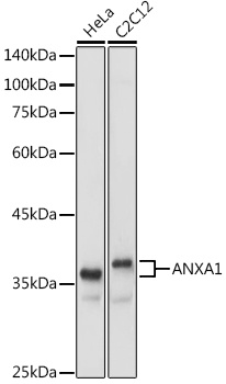
Western blot analysis of various lysates using ANXA1 Rabbit pAb (CAB0767) at 1:1000 dilution. Secondary antibody: HRP-conjugated Goat anti-Rabbit IgG (H+L) (CABS014) at 1:10000 dilution. Lysates/proteins: 25μg per lane. Blocking buffer: 3% nonfat dry milk in TBST. Detection: ECL Basic Kit (AbGn00020). Exposure time: 1s.
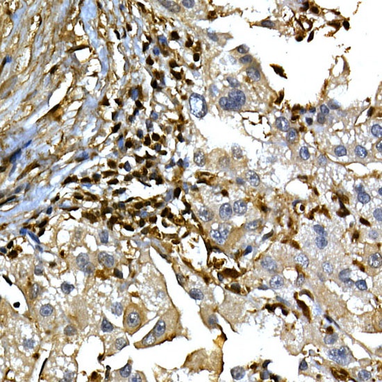
Immunohistochemistry analysis of paraffin-embedded Human liver cancer using ANXA1 Rabbit pAb (CAB0767) at dilution of 1:100 (40x lens). High pressure antigen retrieval performed with 0.01M Citrate buffer (pH 6.0) prior to IHC staining.
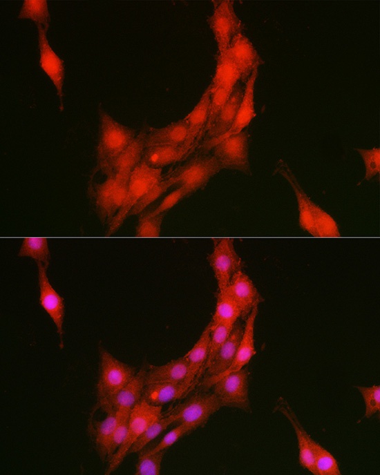
Immunofluorescence analysis of PC-12 cells using ANXA1 Rabbit pAb (CAB0767) at dilution of 1:100 (40x lens). Secondary antibody: Cy3-conjugated Goat anti-Rabbit IgG (H+L) (CABS007) at 1:500 dilution. Blue: DAPI for nuclear staining.
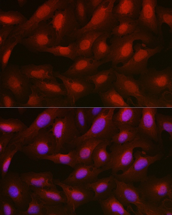
Immunofluorescence analysis of U2OS cells using ANXA1 Rabbit pAb (CAB0767) at dilution of 1:100 (40x lens). Secondary antibody: Cy3-conjugated Goat anti-Rabbit IgG (H+L) (CABS007) at 1:500 dilution. Blue: DAPI for nuclear staining.
 at 1:1000 dilution. Secondary antibody: HRP Goat Anti-Rabbit IgG (H+L) at 1:10000 dilution. Lysates/proteins: 25μg per lane. Blocking buffer: 3% nonfat dry milk in TBST.")
 at 1:1000 dilution. Secondary antibody: HRP Goat Anti-Rabbit IgG (H+L) at 1:10000 dilution. Lysates/proteins: 25μg per lane. Blocking buffer: 3% nonfat dry milk in TBST.")


