The PEBP1 Antibody (CAB0578) is a high-quality antibody developed for reliable detection and analysis of target proteins. This antibody, raised in rabbits, is highly specific to human PEBP1 and is validated for use in various applications including Western blot and immunohistochemistry.PEBP1 is a multifunctional protein involved in various cellular processes such as cell proliferation, apoptosis, and signal transduction. Its role in regulating these processes makes it a key target for research in cancer biology, neurobiology, and developmental biology.
This antibody is validated for use in WB, IF/ICC, IP, ELISA applications and has demonstrated reactivity against Human, Mouse, Rat samples.
Product Name:
PEBP1 Antibody
SKU:
CAB0578
Size:
20μL, 100μL
Reactivity:
Human, Mouse, Rat
Conjugate:
Unconjugated
Immunogen:
Recombinant protein (or fragment).This information is considered to be commercially sensitive.
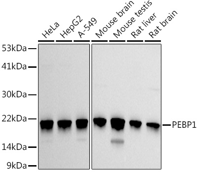
HeLa, HepG2, A-549, Mouse brain, Mouse testis, Rat liver, Rat brain
Cellular Localization:
Cytoplasm.
Calculated MW:
21kDa
Observed MW:
21kDa
This gene encodes a member of the phosphatidylethanolamine-binding family of proteins and has been shown to modulate multiple signaling pathways, including the MAP kinase (MAPK), NF-kappa B, and glycogen synthase kinase-3 (GSK-3) signaling pathways. The encoded protein can be further processed to form a smaller cleavage product, hippocampal cholinergic neurostimulating peptide (HCNP), which may be involved in neural development. This gene has been implicated in numerous human cancers and may act as a metastasis suppressor gene. Multiple pseudogenes of this gene have been identified in the genome.
Purification Method
Affinity purification
Gene ID
5037
RRID
AB_2757275
Buffer Information
Store at -20℃. Avoid freeze / thaw cycles. Buffer: PBS containing 50% glycerol, preserved with proclin300 or sodium azide, pH 7.3.
Western blot analysis of various lysates using PEBP1 Rabbit pAb (CAB0578) at 1:1000 dilution. Secondary antibody: HRP-conjugated Goat anti-Rabbit IgG (H+L) (CABS014) at 1:10000 dilution. Lysates/proteins: 25μg per lane. Blocking buffer: 3% nonfat dry milk in TBST. Detection: ECL Basic Kit (AbGn00020). Exposure time: 30s.
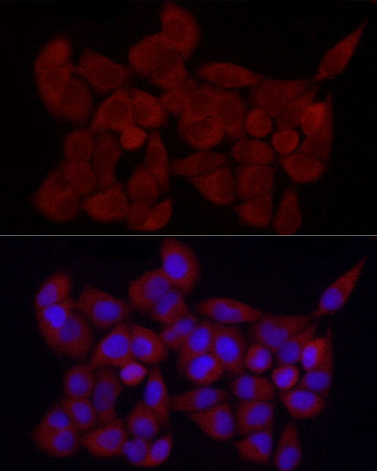
Immunofluorescence analysis of HeLa cells using PEBP1 Rabbit pAb (CAB0578) at dilution of 1:25 (40x lens). Secondary antibody: Cy3-conjugated Goat anti-Rabbit IgG (H+L) (CABS007) at 1:500 dilution. Blue: DAPI for nuclear staining.
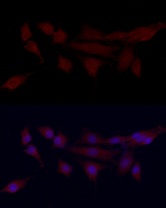
Immunofluorescence analysis of PC-12 cells using PEBP1 Rabbit pAb (CAB0578) at dilution of 1:25 (40x lens). Secondary antibody: Cy3-conjugated Goat anti-Rabbit IgG (H+L) (CABS007) at 1:500 dilution. Blue: DAPI for nuclear staining.
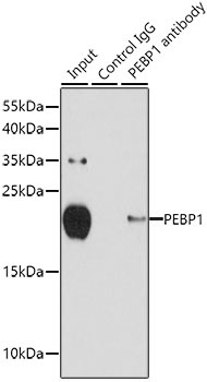
Immunoprecipitation analysis of 150 μg extracts of MCF-7 cells using 3 μg PEBP1 antibody (CAB0578). Western blot was performed from the immunoprecipitate using PEBP1 antibody (CAB0578) at a dilution of 1:500.