The TrkA Antibody (CAB15618) is a high-quality antibody developed for reliable detection and analysis of target proteins. This antibody, produced in rabbits, is specifically designed for use in Western blot applications and is highly reactive with human samples.TrkA is a receptor tyrosine kinase that plays a crucial role in the development and function of the nervous system. Dysregulation of NTRK1 has been implicated in a variety of neurological disorders, including cancer, Alzheimer's disease, and pain syndromes.
This antibody is validated for use in WB, IHC-P, ELISA applications and has demonstrated reactivity against Human, Mouse, Rat samples.
Product Name:
TrkA Antibody
SKU:
CAB15618
Size:
20μL, 100μL
Reactivity:
Human, Mouse, Rat
Conjugate:
Unconjugated
Immunogen:
Synthetic peptide. This information is considered to be commercially sensitive.
Recommended starting concentration is 1 μg/mL. Please optimize the concentration based on your specific assay requirements.
Synonyms:
MTC, TRK, TRK1, TRKA, Trk-A, p140-TrkA, TrkA
Positive Sample:
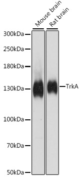
Mouse brain, Rat brain
Cellular Localization:
Cell Membrane, Early Endosome Membrane, Late Endosome Membrane, Single-Pass Type I Membrane Protein.
Calculated MW:
87kDa
Observed MW:
140kDa
This gene encodes a member of the neurotrophic tyrosine kinase receptor (NTKR) family. This kinase is a membrane-bound receptor that, upon neurotrophin binding, phosphorylates itself and members of the MAPK pathway. The presence of this kinase leads to cell differentiation and may play a role in specifying sensory neuron subtypes. Mutations in this gene have been associated with congenital insensitivity to pain, anhidrosis, self-mutilating behavior, cognitive disability and cancer. Alternate transcriptional splice variants of this gene have been found, but only three have been characterized to date.
Purification Method
Affinity purification
Gene ID
4914
RRID
AB_2763024
Buffer Information
Store at -20℃. Avoid freeze / thaw cycles. Buffer: PBS containing 50% glycerol, preserved with proclin300 or sodium azide, pH 7.3.
Western blot analysis of various lysates using TrkA Rabbit pAb (CAB15618) at 1:500 dilution. Secondary antibody: HRP-conjugated Goat anti-Rabbit IgG (H+L) (CABS014) at 1:10000 dilution. Lysates/proteins: 25μg per lane. Blocking buffer: 3% nonfat dry milk in TBST. Detection: ECL Basic Kit (AbGn00020). Exposure time: 90s.
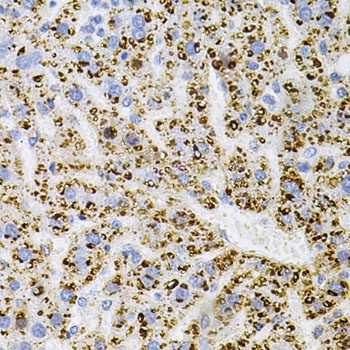
Immunohistochemistry analysis of paraffin-embedded Rat liver using TrkA Rabbit pAb (CAB15618) at dilution of 1:100 (40x lens). Microwave antigen retrieval performed with 0.01M PBS Buffer (pH 7.2) prior to IHC staining.
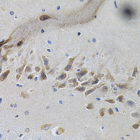
Immunohistochemistry analysis of paraffin-embedded Mouse brain using TrkA Rabbit pAb (CAB15618) at dilution of 1:100 (40x lens). Microwave antigen retrieval performed with 0.01M PBS Buffer (pH 7.2) prior to IHC staining.