The NMNAT1 Antibody (CAB6672) is a high-quality antibody developed for reliable detection and analysis of target proteins. This antibody, generated in rabbits, exhibits high reactivity with human samples and has been validated for use in immunohistochemistry (IHC) and Western blot applications.NMNAT1 is known for its role in maintaining cellular health and promoting neuronal survival, making it a key player in neurodegenerative diseases such as Alzheimer's and Parkinson's.
This antibody is validated for use in WB, ELISA applications and has demonstrated reactivity against Human samples.
Product Name:
NMNAT1 Antibody
SKU:
CAB6672
Size:
20μL, 100μL
Reactivity:
Human
Conjugate:
Unconjugated
Immunogen:
Recombinant protein (or fragment).This information is considered to be commercially sensitive.
Recommended starting concentration is 1 μg/mL. Please optimize the concentration based on your specific assay requirements.
Synonyms:
LCA9, NMNAT, PNAT1, SHILCA, NMNAT1
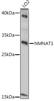
Positive Sample:
LO2
Cellular Localization:
Nucleus.
Calculated MW:
32kDa
Observed MW:
32kDa
This gene encodes an enzyme which catalyzes a key step in the biosynthesis of nicotinamide adenine dinucleotide (NAD). The encoded enzyme is one of several nicotinamide nucleotide adenylyltransferases, and is specifically localized to the cell nucleus. Activity of this protein leads to the activation of a nuclear deacetylase that functions in the protection of damaged neurons. Mutations in this gene have been associated with Leber congenital amaurosis 9. Alternative splicing results in multiple transcript variants. Pseudogenes of this gene are located on chromosomes 1, 3, 4, 14, and 15.
Purification Method
Affinity purification
Gene ID
64802
RRID
AB_2767257
Buffer Information
Store at -20℃. Avoid freeze / thaw cycles. Buffer: PBS containing 50% glycerol, preserved with proclin300 or sodium azide, pH 7.3.
Western blot analysis of lysates from LO2 cells, using NMNAT1 Rabbit pAb (CAB6672) at 1:1000 dilution. Secondary antibody: HRP-conjugated Goat anti-Rabbit IgG (H+L) (CABS014) at 1:10000 dilution. Lysates/proteins: 25μg per lane. Blocking buffer: 3% nonfat dry milk in TBST. Detection: ECL Basic Kit (AbGn00020). Exposure time: 30s.