The NEK8 Antibody (CAB0984) is a high-quality antibody developed for reliable detection and analysis of target proteins. The antibody, generated in rabbits, exhibits high specificity and sensitivity towards human samples, making it ideal for use in Western blotting and immunohistochemistry applications.NEK8 is known for its involvement in various cellular processes, including cell division, DNA damage response, and ciliary function. Dysfunction of NEK8 has been linked to ciliopathies, a group of genetic disorders characterized by abnormal cilia structure and function.
This antibody is validated for use in WB, IF/ICC, ELISA applications and has demonstrated reactivity against Human, Mouse, Rat samples.
Product Name:
NEK8 Antibody
SKU:
CAB0984
Size:
20μL, 100μL
Reactivity:
Human, Mouse, Rat
Conjugate:
Unconjugated
Immunogen:
Recombinant protein (or fragment).This information is considered to be commercially sensitive.
Recommended starting concentration is 1 μg/mL. Please optimize the concentration based on your specific assay requirements.
Synonyms:
JCK, NPHP9, RHPD2, NEK12A, NEK8
Positive Sample:
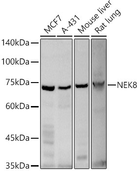
MCF7, A-431, Mouse liver, Rat lung,
Cellular Localization:
Cell Projection, Cytoplasm, Cilium, Cytoskeleton.
Calculated MW:
75kDa
Observed MW:
70kDa
This gene encodes a member of the serine/threionine protein kinase family related to NIMA (never in mitosis, gene A) of Aspergillus nidulans. The encoded protein may play a role in cell cycle progression from G2 to M phase. Mutations in the related mouse gene are associated with a disease phenotype that closely parallels the juvenile autosomal recessive form of polycystic kidney disease in humans.
Purification Method
Affinity purification
Gene ID
284086
RRID
AB_2757503
Buffer Information
Store at -20℃. Avoid freeze / thaw cycles. Buffer: PBS containing 50% glycerol, preserved with proclin300 or sodium azide, pH 7.3.
Western blot analysis of various lysates using NEK8 Rabbit pAb (CAB0984) at 1:500 dilution. Secondary antibody: HRP-conjugated Goat anti-Rabbit IgG (H+L) (CABS014) at 1:10000 dilution. Lysates / proteins: 25 μg per lane. Blocking buffer: 3 % nonfat dry milk in TBST. Detection: ECL Basic Kit (AbGn00020). Exposure time: 180s.
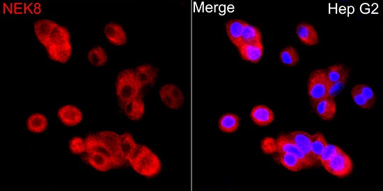
Immunofluorescence analysis of HepG2 cells using NEK8 Rabbit pAb (CAB0984) at dilution of 1:500 (40x lens). Secondary antibody: Cy3-conjugated Goat anti-Rabbit IgG (H+L) (CABS007) at 1:500 dilution. Blue: DAPI for nuclear staining.