The NDUFV1 Antibody (CAB8014) is a high-quality antibody developed for reliable detection and analysis of target proteins. This antibody, generated in rabbits, demonstrates high specificity for human samples and is optimized for use in Western blot analysis. By binding to NDUFAF1, it enables precise detection and analysis of this protein in a variety of cell types, making it ideal for investigations in mitochondrial biology and metabolic diseases.NDUFAF1 is a crucial factor in the assembly of complex I of the mitochondrial respiratory chain, essential for ATP production and cellular energy metabolism.
This antibody is validated for use in WB, IHC-P, ELISA applications and has demonstrated reactivity against Human, Mouse, Rat samples.
Product Name:
NDUFV1 Antibody
SKU:
CAB8014
Size:
20μL, 100μL
Reactivity:
Human, Mouse, Rat
Conjugate:
Unconjugated
Immunogen:
Recombinant protein (or fragment).This information is considered to be commercially sensitive.
The mitochondrial respiratory chain provides energy to cells via oxidative phosphorylation and consists of four membrane-bound electron-transporting protein complexes (I-IV) and an ATP synthase (complex V). This gene encodes a 51 kDa subunit of the NADH:ubiquinone oxidoreductase complex I; a large complex with at least 45 nuclear and mitochondrial encoded subunits that liberates electrons from NADH and channels them to ubiquinone. This subunit carries the NADH-binding site as well as flavin mononucleotide (FMN)- and Fe-S-biding sites. Defects in complex I are a common cause of mitochondrial dysfunction; a syndrome that occurs in approximately 1 in 10,000 live births. Mitochondrial complex I deficiency is linked to myopathies, encephalomyopathies, and neurodegenerative disorders such as Parkinson's disease and Leigh syndrome. Alternative splicing results in multiple transcript variants encoding distinct isoforms.
Purification Method
Affinity purification
Gene ID
4723
RRID
AB_2770553
Buffer Information
Store at -20℃. Avoid freeze / thaw cycles. Buffer: PBS containing 50% glycerol, preserved with proclin300 or sodium azide, pH 7.3.
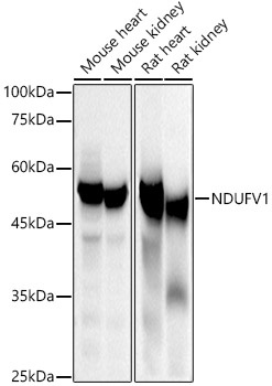
Western blot analysis of various lysates using NDUFV1 Rabbit pAb (CAB8014) at 1:500 dilution. Secondary antibody: HRP-conjugated Goat anti-Rabbit IgG (H+L) (CABS014) at 1:10000 dilution. Lysates/proteins: 25μg per lane. Blocking buffer: 3% nonfat dry milk in TBST. Detection: ECL Basic Kit (AbGn00020). Exposure time: 10s.
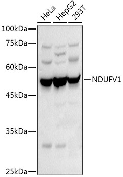
Western blot analysis of various lysates using NDUFV1 Rabbit pAb (CAB8014) at 1:500 dilution. Secondary antibody: HRP-conjugated Goat anti-Rabbit IgG (H+L) (CABS014) at 1:10000 dilution. Lysates/proteins: 25μg per lane. Blocking buffer: 3% nonfat dry milk in TBST. Detection: ECL Basic Kit (AbGn00020). Exposure time: 30s.
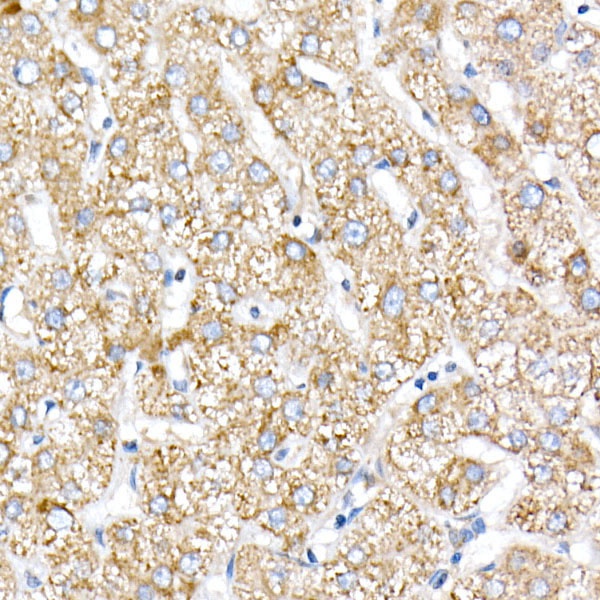
Immunohistochemistry analysis of paraffin-embedded Human liver using NDUFV1 Rabbit pAb (CAB8014) at dilution of 1:20 (40x lens). High pressure antigen retrieval performed with 0.01M Citrate buffer (pH 6.0) prior to IHC staining.
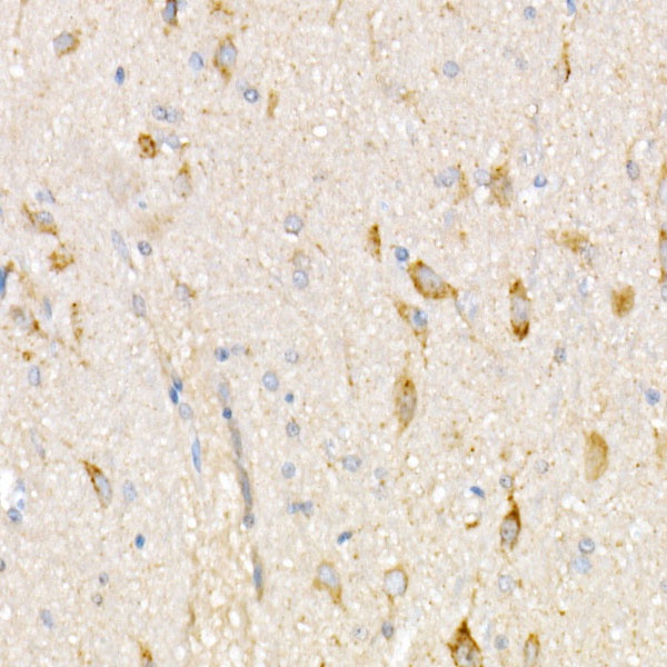
Immunohistochemistry analysis of paraffin-embedded Rat brain using NDUFV1 Rabbit pAb (CAB8014) at dilution of 1:20 (40x lens). High pressure antigen retrieval performed with 0.01M Citrate buffer (pH 6.0) prior to IHC staining.