The MSP/MST1 Antibody (CAB12963) is a high-quality antibody developed for reliable detection and analysis of target proteins. This antibody is raised in rabbits and is highly reactive with human samples, making it ideal for use in Western blot applications. The MST1 Polyclonal Antibody binds specifically to the MST1 protein, allowing for precise detection and analysis in various cell types.MST1, also known as mammalian sterile 20-like kinase 1, is involved in multiple cellular processes, including cell growth, differentiation, and survival. Dysregulation of MST1 has been implicated in various diseases, including cancer, cardiovascular disorders, and neurodegenerative conditions.
This antibody is validated for use in WB, IHC-P, IF/ICC, ELISA applications and has demonstrated reactivity against Human, Mouse, Rat samples.
Product Name:
MSP/MST1 Antibody
SKU:
CAB12963
Size:
20μL, 100μL
Reactivity:
Human, Mouse, Rat
Conjugate:
Unconjugated
Immunogen:
Recombinant protein (or fragment).This information is considered to be commercially sensitive.
Recommended starting concentration is 1 μg/mL. Please optimize the concentration based on your specific assay requirements.
Synonyms:
MSP, HGFL, NF15S2, D3F15S2, DNF15S2, MSP/MST1
Positive Sample:
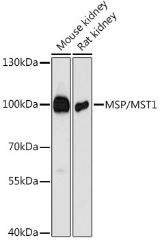
Mouse kidney, Rat kidney
Cellular Localization:
Secreted.
Calculated MW:
80kDa
Observed MW:
100kDa
The protein encoded by this gene contains four kringle domains and a serine protease domain, similar to that found in hepatic growth factor. Despite the presence of the serine protease domain, the encoded protein may not have any proteolytic activity. The receptor for this protein is RON tyrosine kinase, which upon activation stimulates ciliary motility of ciliated epithelial lung cells. This protein is secreted and cleaved to form an alpha chain and a beta chain bridged by disulfide bonds.
Purification Method
Affinity purification
Gene ID
4485
RRID
AB_2759810
Buffer Information
Store at -20℃. Avoid freeze / thaw cycles. Buffer: PBS with 0.01% thimerosal,50% glycerol,pH7.3.
Western blot analysis of various lysates using MSP/MST1 Rabbit pAb (CAB12963) at 1:3000 dilution. Secondary antibody: HRP-conjugated Goat anti-Rabbit IgG (H+L) (CABS014) at 1:10000 dilution. Lysates/proteins: 25μg per lane. Blocking buffer: 3% nonfat dry milk in TBST. Detection: ECL Basic Kit (AbGn00020). Exposure time: 90s.
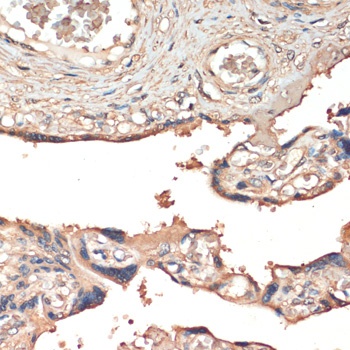
Immunohistochemistry analysis of paraffin-embedded Human placenta using MSP/MST1 Rabbit pAb (CAB12963) at dilution of 1:200 (40x lens). Microwave antigen retrieval performed with 0.01M PBS Buffer (pH 7.2) prior to IHC staining.
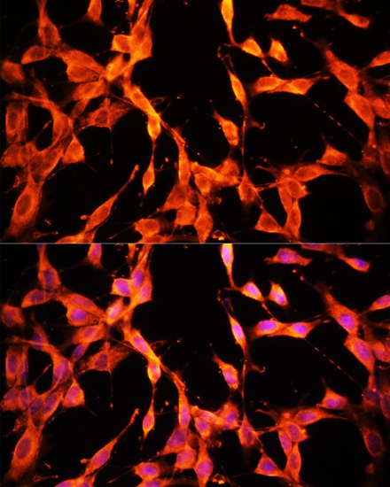
Immunofluorescence analysis of NIH/3T3 cells using MSP/MST1 Rabbit pAb (CAB12963) at dilution of 1:100. Secondary antibody: Cy3-conjugated Goat anti-Rabbit IgG (H+L) (CABS007) at 1:500 dilution. Blue: DAPI for nuclear staining.