The MLH3 Antibody (CAB7796) is a high-quality antibody developed for reliable detection and analysis of target proteins. This antibody, raised in rabbits, shows high reactivity with human samples and has been validated for use in Western blot applications. By binding specifically to the MLH3 protein, this antibody enables precise detection and analysis in a variety of cell types, making it an ideal choice for studies in genetics, cancer research, and molecular biology.MLH3 is a crucial component of the mismatch repair system, which plays a vital role in maintaining genomic stability and preventing mutations that can lead to cancer and other genetic disorders.
This antibody is validated for use in WB, ELISA applications and has demonstrated reactivity against Human, Mouse, Rat samples.
Product Name:
MLH3 Antibody
SKU:
CAB7796
Size:
20μL, 100μL
Reactivity:
Human, Mouse, Rat
Conjugate:
Unconjugated
Immunogen:
Recombinant protein (or fragment).This information is considered to be commercially sensitive.
Recommended starting concentration is 1 μg/mL. Please optimize the concentration based on your specific assay requirements.
Synonyms:
HNPCC7, MLH3
Positive Sample:
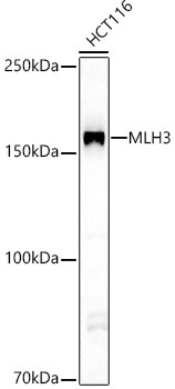
HCT116
Cellular Localization:
Nucleus.
Calculated MW:
164kDa
Observed MW:
164kDa
This gene is a member of the MutL-homolog (MLH) family of DNA mismatch repair (MMR) genes. MLH genes are implicated in maintaining genomic integrity during DNA replication and after meiotic recombination. The protein encoded by this gene functions as a heterodimer with other family members. Somatic mutations in this gene frequently occur in tumors exhibiting microsatellite instability, and germline mutations have been linked to hereditary nonpolyposis colorectal cancer type 7 (HNPCC7). Several alternatively spliced transcript variants have been identified, but the full-length nature of only two transcript variants has been determined.
Purification Method
Affinity purification
Gene ID
27030
RRID
AB_2770374
Buffer Information
Store at -20℃. Avoid freeze / thaw cycles. Buffer: PBS containing 50% glycerol, preserved with proclin300 or sodium azide, pH 7.3.
Western blot analysis of lysates from HCT116 cells, using MLH3 Rabbit pAb (CAB7796) at 1:3500 dilution. Secondary antibody: HRP-conjugated Goat anti-Rabbit IgG (H+L) (CABS014) at 1:10000 dilution. Lysates/proteins: 25μg per lane. Blocking buffer: 3% nonfat dry milk in TBST. Detection: ECL Basic Kit (AbGn00020). Exposure time: 60s.