The LRRC59 Antibody (CAB10022) is a high-quality antibody developed for reliable detection and analysis of target proteins. This antibody is produced in rabbits and exhibits high reactivity with human samples, making it a reliable tool for immunodetection assays like Western blotting.LRRC59, also known as Leucine-Rich Repeat Containing 59, is involved in protein-protein interactions and has been linked to cell signaling pathways and gene expression regulation.
This antibody is validated for use in WB, IHC-P, ELISA applications and has demonstrated reactivity against Human, Mouse, Rat samples.
Product Name:
LRRC59 Antibody
SKU:
CAB10022
Size:
20μL, 100μL
Reactivity:
Human, Mouse, Rat
Conjugate:
Unconjugated
Immunogen:
Recombinant protein (or fragment).This information is considered to be commercially sensitive.
Recommended starting concentration is 1 μg/mL. Please optimize the concentration based on your specific assay requirements.
Synonyms:
p34, PRO1855, 59
Positive Sample:
293T
Cellular Localization:
Endoplasmic Reticulum Membrane, Microsome Membrane, Nucleus Envelope, Single-Pass Type Ii Membrane Protein.
Calculated MW:
35kDa
Observed MW:
35kDa
Enables RNA binding activity and cadherin binding activity. Predicted to be involved in positive regulation of Ras protein signal transduction and signal transduction. Located in endoplasmic reticulum and mitochondrial nucleoid.
Purification Method
Affinity purification
Gene ID
55379
RRID
AB_2757542
Buffer Information
Store at -20℃. Avoid freeze / thaw cycles. Buffer: PBS containing 50% glycerol, preserved with proclin300 or sodium azide, pH 7.3.
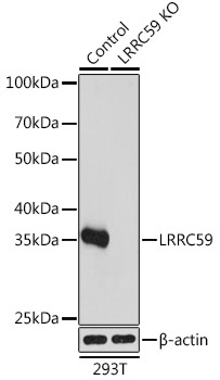
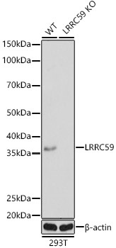
Western blot analysis of lysates from wild type (WT) and LRRC59 knockout (KO) 293T cells, using [KO Validated] LRRC59 Rabbit pAb (CAB10022) at 1:1000 dilution. Secondary antibody: HRP-conjugated Goat anti-Rabbit IgG (H+L) (CABS014) at 1:10000 dilution. Lysates/proteins: 25μg per lane. Blocking buffer: 3% nonfat dry milk in TBST. Detection: ECL Basic Kit (AbGn00020). Exposure time: 1s.
Western blot analysis of lysates from wild type(WT) and LRRC59 knockout (KO) 293T(KO) cells, using [KO Validated] LRRC59 Rabbit pAb (CAB10022) at 1:1000 dilution. Secondary antibody: HRP-conjugated Goat anti-Rabbit IgG (H+L) (CABS014) at 1:10000 dilution. Lysates/proteins: 25μg per lane. Blocking buffer: 3% nonfat dry milk in TBST. Detection: ECL Basic Kit (AbGn00020). Exposure time: 1s.
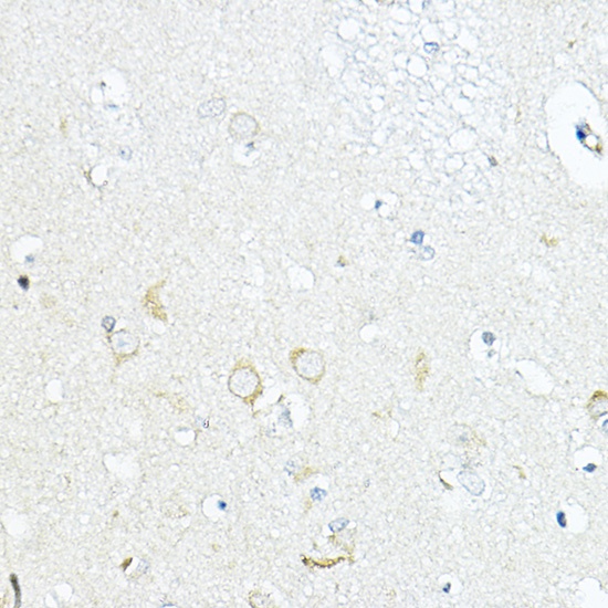
Immunohistochemistry analysis of paraffin-embedded Human brain using LRRC59 Rabbit pAb (CAB10022) at dilution of 1:100 (40x lens). High pressure antigen retrieval performed with 0.01M Citrate buffer (pH 6.0) prior to IHC staining.
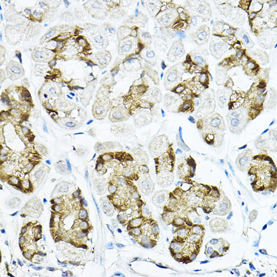
Immunohistochemistry analysis of paraffin-embedded Mouse stomach using LRRC59 Rabbit pAb (CAB10022) at dilution of 1:100 (40x lens). High pressure antigen retrieval performed with 0.01M Citrate buffer (pH 6.0) prior to IHC staining.