The [KO Validated] Smad3 Monoclonal Antibody (CAB19115) is a high-quality antibody developed for reliable detection and analysis of target proteins. This antibody, raised in rabbits, has been validated for use in techniques such as Western blot and immunohistochemistry, providing reliable detection and analysis of SMAD3 protein expression in human samples.SMAD3 is a key transcription factor that plays a crucial role in mediating TGF-β signaling, which is important for cellular responses such as cell proliferation, differentiation, and migration.
This antibody is validated for use in WB, IHC-P, IP, ChIP, ELISA applications and has demonstrated reactivity against Human, Mouse, Rat samples.
Product Name:
[KO Validated] Smad3 Monoclonal Antibody
SKU:
CAB19115
Size:
20μL, 100μL
Reactivity:
Human, Mouse, Rat
Clone Number:
ARC53861
Conjugate:
Unconjugated
Immunogen:
Synthetic peptide. This information is considered to be commercially sensitive.
The SMAD family of proteins are a group of intracellular signal transducer proteins similar to the gene products of the Drosophila gene 'mothers against decapentaplegic' (Mad) and the C. elegans gene Sma. The SMAD3 protein functions in the transforming growth factor-beta signaling pathway, and transmits signals from the cell surface to the nucleus, regulating gene activity and cell proliferation. This protein forms a complex with other SMAD proteins and binds DNA, functioning both as a transcription factor and tumor suppressor. Mutations in this gene are associated with aneurysms-osteoarthritis syndrome and Loeys-Dietz Syndrome 3.
Purification Method
Protein A
Gene ID
4088
RRID
AB_2862608
Buffer Information
Store at -20℃. Avoid freeze / thaw cycles. Buffer: PBS containing 50% glycerol and 0.05% BSA, preserved with proclin300 or sodium azide, pH 7.3.
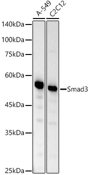
Western blot analysis of various lysates, using Smad3 Rabbit mAb (CAB19115) at 1:20000 dilution. Secondary antibody: HRP-conjugated Goat anti-Rabbit IgG (H+L) (CABS014) at 1:10000 dilution. Lysates/proteins: 25μg per lane. Blocking buffer: 3% nonfat dry milk in TBST. Detection: ECL Basic Kit (AbGn00020). Exposure time: 30s.
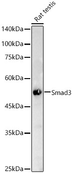
Western blot analysis of lysates from Rat testis, using Smad3 Rabbit mAb (CAB19115) at 1:20000 dilution. Secondary antibody: HRP-conjugated Goat anti-Rabbit IgG (H+L) (CABS014) at 1:10000 dilution. Lysates/proteins: 25μg per lane. Blocking buffer: 3% nonfat dry milk in TBST. Detection: ECL Basic Kit (AbGn00020). Exposure time: 30s.
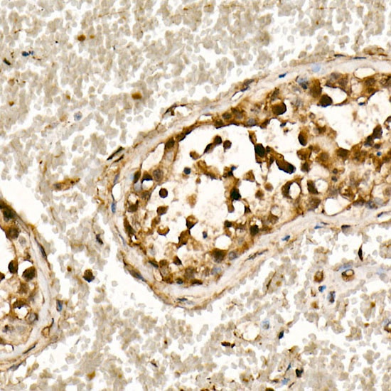
Immunohistochemistry analysis of paraffin-embedded Human testis using [KO Validated] Smad3 Rabbit mAb (CAB19115) at dilution of 1:200 (40x lens). High pressure antigen retrieval performed with 0.01M Citrate buffer (pH 6.0) prior to IHC staining.
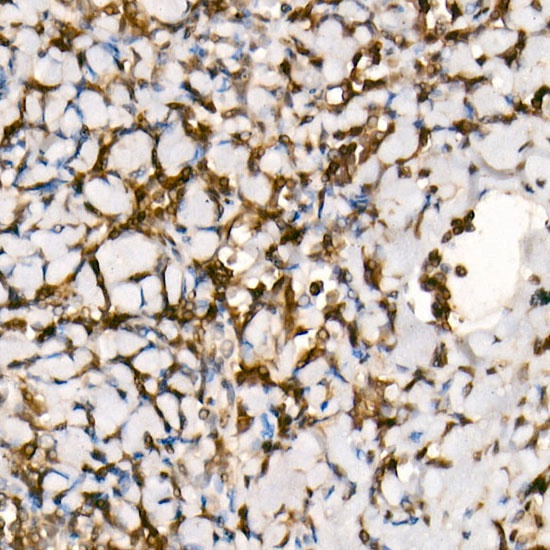
Immunohistochemistry analysis of paraffin-embedded Human tonsil using [KO Validated] Smad3 Rabbit mAb (CAB19115) at dilution of 1:200 (40x lens). High pressure antigen retrieval performed with 0.01M Citrate buffer (pH 6.0) prior to IHC staining.