The KLK10 Antibody (CAB6398) is a high-quality antibody developed for reliable detection and analysis of target proteins. This antibody, raised in rabbits, exhibits high reactivity with human samples and has been validated for use in Western blot applications. By specifically binding to Kallikrein 10, researchers can detect and analyze this protein in different cell types, making it an ideal choice for studies in cancer research and biomarker discovery.
This antibody is validated for use in WB, IF/ICC, ELISA applications and has demonstrated reactivity against Human, Mouse, Rat samples.
Product Name:
KLK10 Antibody
SKU:
CAB6398
Size:
20μL, 100μL
Reactivity:
Human, Mouse, Rat
Conjugate:
Unconjugated
Immunogen:
Recombinant protein (or fragment).This information is considered to be commercially sensitive.
Recommended starting concentration is 1 μg/mL. Please optimize the concentration based on your specific assay requirements.
Synonyms:
NES1, PRSSL1, KLK10
Positive Sample:
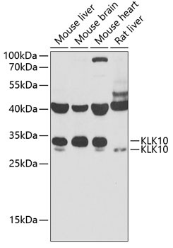
Mouse liver, Mouse brain, Mouse heart, Rat liver
Cellular Localization:
Secreted.
Calculated MW:
30kDa
Observed MW:
30kDa
Kallikreins are a subgroup of serine proteases having diverse physiological functions. Growing evidence suggests that many kallikreins are implicated in carcinogenesis and some have potential as novel cancer and other disease biomarkers. This gene is one of the fifteen kallikrein subfamily members located in a cluster on chromosome 19. Its encoded protein is secreted and may play a role in suppression of tumorigenesis in breast and prostate cancers. Alternate splicing of this gene results in multiple transcript variants encoding the same protein.
Purification Method
Affinity purification
Gene ID
5655
RRID
AB_2767000
Buffer Information
Store at -20℃. Avoid freeze / thaw cycles. Buffer: PBS containing 50% glycerol, preserved with proclin300 or sodium azide, pH 7.3.
Western blot analysis of various lysates using KLK10 Rabbit pAb (CAB6398) at 1:1000 dilution. Secondary antibody: HRP-conjugated Goat anti-Rabbit IgG (H+L) (CABS014) at 1:10000 dilution. Lysates/proteins: 25μg per lane. Blocking buffer: 3% nonfat dry milk in TBST. Detection: ECL Basic Kit (AbGn00020). Exposure time: 90s.
Immunofluorescence analysis of MCF7 cells using KLK10 Rabbit pAb (CAB6398) at dilution of 1:400 (40x lens). Secondary antibody: Cy3-conjugated Goat anti-Rabbit IgG (H+L) (CABS007) at 1:500 dilution. Blue: DAPI for nuclear staining.
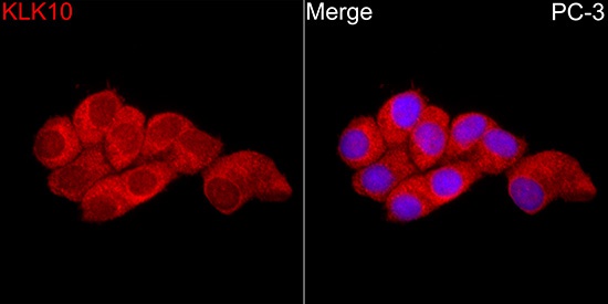
Immunofluorescence analysis of PC-3 cells using KLK10 Rabbit pAb (CAB6398) at dilution of 1:400 (40x lens). Secondary antibody: Cy3-conjugated Goat anti-Rabbit IgG (H+L) (CABS007) at 1:500 dilution. Blue: DAPI for nuclear staining.