The IRF1 Antibody (CAB7692) is a high-quality antibody developed for reliable detection and analysis of target proteins. This antibody, raised in rabbits, exhibits high reactivity with human samples and has been validated for use in Western blot applications. By binding specifically to IRF1, researchers can effectively detect and analyze this important protein in various cell types.IRF1 is a key transcription factor involved in regulating immune responses, playing a crucial role in the activation of immune cells and the expression of genes involved in anti-viral and anti-tumor responses.
This antibody is validated for use in WB, IHC-P, IF/ICC, ELISA applications and has demonstrated reactivity against Human, Mouse, Rat samples.
Product Name:
IRF1 Antibody
SKU:
CAB7692
Size:
20μL, 100μL
Reactivity:
Human, Mouse, Rat
Conjugate:
Unconjugated
Immunogen:
Synthetic peptide. This information is considered to be commercially sensitive.
Recommended starting concentration is 1 μg/mL. Please optimize the concentration based on your specific assay requirements.
Synonyms:
MAR, IRF-1, IRF1
Positive Sample:
Rat spleen, C2C12 treated with IFNγ
Cellular Localization:
Cytoplasm, Nucleus.
Calculated MW:
37kDa
Observed MW:
48kDa
The protein encoded by this gene is a transcriptional regulator and tumor suppressor, serving as an activator of genes involved in both innate and acquired immune responses. The encoded protein activates the transcription of genes involved in the body's response to viruses and bacteria, playing a role in cell proliferation, apoptosis, the immune response, and DNA damage response. This protein represses the transcription of several other genes. As a tumor suppressor, it both suppresses tumor cell growth and stimulates an immune response against tumor cells. Defects in this gene have been associated with gastric cancer, myelogenous leukemia, and lung cancer.
Purification Method
Affinity purification
Gene ID
3659
RRID
AB_2769990
Buffer Information
Store at -20℃. Avoid freeze / thaw cycles. Buffer: PBS containing 50% glycerol, preserved with proclin300 or sodium azide, pH 7.3.
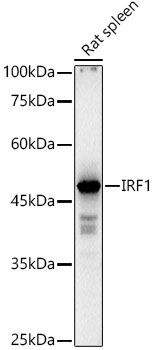
Western blot analysis of lysates from Rat spleen, using IRF1 Rabbit pAb (CAB7692) at 1:1000 dilution. Secondary antibody: HRP-conjugated Goat anti-Rabbit IgG (H+L) (CABS014) at 1:10000 dilution. Lysates/proteins: 25μg per lane. Blocking buffer: 3% nonfat dry milk in TBST. Detection: ECL Basic Kit (AbGn00020). Exposure time: 10s.
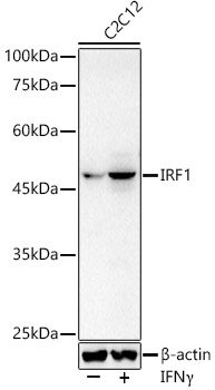
Western blot analysis of lysates from C2C12 cells, using IRF1 Rabbit pAb (CAB7692) at 1:1000 dilution. C2C12 cells were treated with IFNγ (50ng/ml) for 8 hours. Secondary antibody: HRP-conjugated Goat anti-Rabbit IgG (H+L) (CABS014) at 1:10000 dilution. Lysates/proteins: 25μg per lane. Blocking buffer: 3% nonfat dry milk in TBST. Detection: ECL Basic Kit (AbGn00020). Exposure time: 10s.
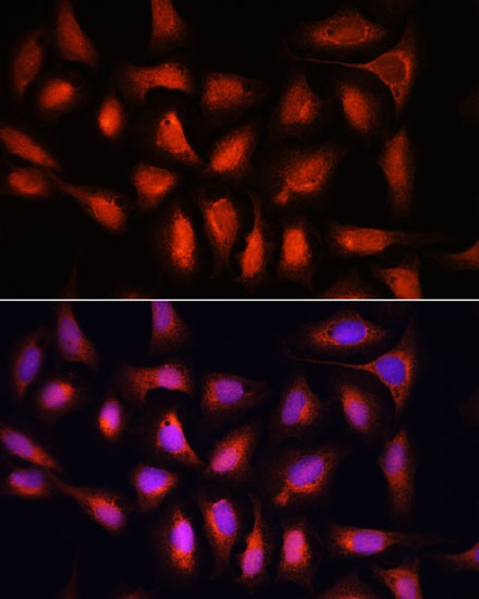
Immunofluorescence analysis of U-2 OS cells using IRF1 Rabbit pAb (CAB7692) at dilution of 1:100. Secondary antibody: Cy3-conjugated Goat anti-Rabbit IgG (H+L) (CABS007) at 1:500 dilution. Blue: DAPI for nuclear staining.