The HINT1 Polyclonal Antibody (CAB21797) is a high-quality antibody developed for reliable detection and analysis of target proteins. This antibody, produced in rabbits, is highly specific to human samples and is validated for use in Western blot applications. By binding to the HINT1 protein, this antibody enables the detection and analysis of HINT1 in a wide range of cell types, making it essential for studies in biochemistry and molecular biology.
This antibody is validated for use in WB, IF/ICC, ELISA applications and has demonstrated reactivity against Human, Mouse, Rat samples.
Product Name:
HINT1 Polyclonal Antibody
SKU:
CAB21797
Size:
20μL, 100μL
Reactivity:
Human, Mouse, Rat
Conjugate:
Unconjugated
Immunogen:
Recombinant protein (or fragment).This information is considered to be commercially sensitive.
Recommended starting concentration is 1 μg/mL. Please optimize the concentration based on your specific assay requirements.
Synonyms:
HINT, NMAN, PKCI-1, PRKCNH1, HINT1
Positive Sample:
Hela, H460, NIH/3T3
Cellular Localization:
Cytoplasm, Nucleus.
Calculated MW:
14kDa
Observed MW:
Refertofigures
This gene encodes a protein that hydrolyzes purine nucleotide phosphoramidates substrates, including AMP-morpholidate, AMP-N-alanine methyl ester, AMP-alpha-acetyl lysine methyl ester, and AMP-NH2. The encoded protein interacts with these substrates via a histidine triad motif. This gene is considered a tumor suppressor gene. In addition, mutations in this gene can cause autosomal recessive neuromyotonia and axonal neuropathy. There are several related pseudogenes on chromosome 7. Several transcript variants have been observed.
Purification Method
Affinity purification
Gene ID
3094
Buffer Information
Store at -20℃. Avoid freeze / thaw cycles. Buffer: PBS containing 50% glycerol, preserved with proclin300 or sodium azide, pH 7.3.
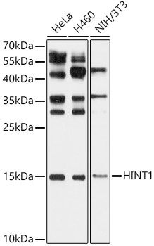
Western blot analysis of various lysates, using HINT1 Rabbit pAb (CAB21797) at 1:1000 dilution. Secondary antibody: HRP-conjugated Goat anti-Rabbit IgG (H+L) (CABS014) at 1:10000 dilution. Lysates/proteins: 25μg per lane. Blocking buffer: 3% nonfat dry milk in TBST. Detection: ECL Basic Kit (AbGn00020). Exposure time: 5s.
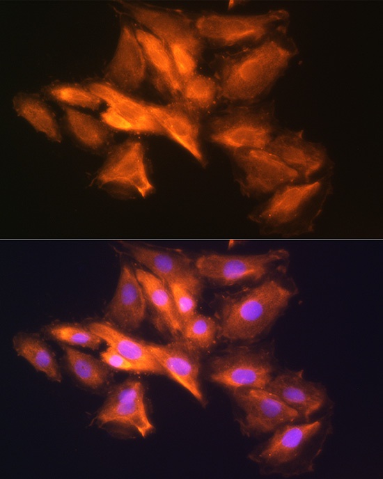
Immunofluorescence analysis of H9C2 cells using HINT1 Rabbit pAb (CAB21797) at dilution of 1:100 (40x lens). Secondary antibody: Cy3-conjugated Goat anti-Rabbit IgG (H+L) (CABS007) at 1:500 dilution. Blue: DAPI for nuclear staining.
 at 1:1000 dilution. Secondary antibody: HRP Goat Anti-Rabbit IgG (H+L) at 1:10000 dilution. Lysates/proteins: 25μg per lane. Blocking buffer: 3% nonfat dry milk in TBST.")
 at 1:1000 dilution. Secondary antibody: HRP Goat Anti-Rabbit IgG (H+L) at 1:10000 dilution. Lysates/proteins: 25μg per lane. Blocking buffer: 3% nonfat dry milk in TBST.")


