The HDGF Antibody (CAB5347) is a high-quality antibody developed for reliable detection and analysis of target proteins. This antibody, produced in rabbits, exhibits high specificity towards HDGF in human samples and is validated for use in various applications, including Western blot and immunohistochemistry.HDGF is known to play a crucial role in promoting cell proliferation, migration, and survival in various cancer types, making it a potential target for cancer therapy.
This antibody is validated for use in WB, IHC-P, IF/ICC, ELISA applications and has demonstrated reactivity against Human, Mouse, Rat samples.
Product Name:
HDGF Antibody
SKU:
CAB5347
Size:
20μL, 100μL
Reactivity:
Human, Mouse, Rat
Conjugate:
Unconjugated
Immunogen:
Recombinant protein (or fragment).This information is considered to be commercially sensitive.
Recommended starting concentration is 1 μg/mL. Please optimize the concentration based on your specific assay requirements.
Synonyms:
HMG1L2, HDGF
Positive Sample:
22Rv1, HeLa, A-549, 293T, U-251MG, MCF7, BT-474
Cellular Localization:
Cytoplasm, Nucleus.
Calculated MW:
27kDa
Observed MW:
38kDa
This gene encodes a member of the hepatoma-derived growth factor family. The encoded protein has mitogenic and DNA-binding activity and may play a role in cellular proliferation and differentiation. High levels of expression of this gene enhance the growth of many tumors. This gene was thought initially to be located on chromosome X; however, that location has been determined to correspond to a related pseudogene. Alternatively spliced transcript variants encoding distinct isoforms have been described.
Purification Method
Affinity purification
Gene ID
3068
RRID
AB_2766157
Buffer Information
Store at -20℃. Avoid freeze / thaw cycles. Buffer: PBS containing 50% glycerol, preserved with proclin300 or sodium azide, pH 7.3.
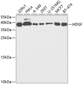
Western blot analysis of various lysates using HDGF Rabbit pAb (CAB5347) at 1:1000 dilution. Secondary antibody: HRP-conjugated Goat anti-Rabbit IgG (H+L) (CABS014) at 1:10000 dilution. Lysates/proteins: 25μg per lane. Blocking buffer: 3% nonfat dry milk in TBST. Detection: ECL Basic Kit (AbGn00020). Exposure time: 90s.
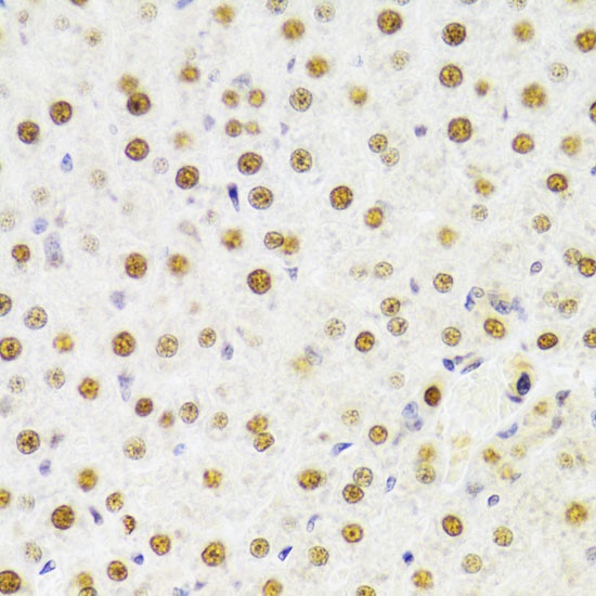
Immunohistochemistry analysis of paraffin-embedded Rat liver using HDGF Rabbit pAb (CAB5347) at dilution of 1:100 (40x lens). Microwave antigen retrieval performed with 0.01M PBS Buffer (pH 7.2) prior to IHC staining.
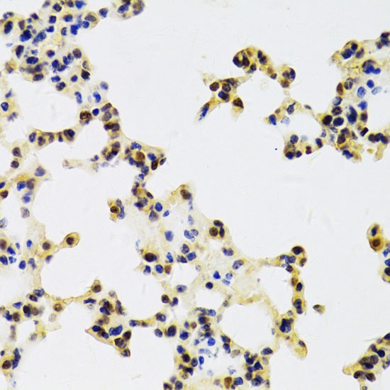
Immunohistochemistry analysis of paraffin-embedded Mouse lung using HDGF Rabbit pAb (CAB5347) at dilution of 1:100 (40x lens). Microwave antigen retrieval performed with 0.01M PBS Buffer (pH 7.2) prior to IHC staining.
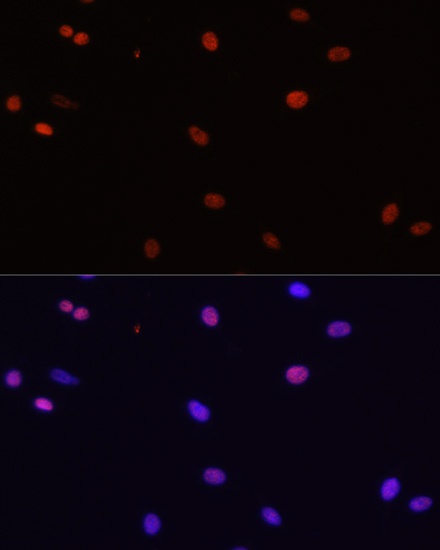
Immunofluorescence analysis of C6 cells using HDGF Rabbit pAb (CAB5347) at dilution of 1:100. Secondary antibody: Cy3-conjugated Goat anti-Rabbit IgG (H+L) (CABS007) at 1:500 dilution. Blue: DAPI for nuclear staining.