The GLDC Polyclonal Antibody (CAB24541) is a high-quality antibody developed for reliable detection and analysis of target proteins. This antibody, generated in rabbits, exhibits high reactivity with human samples and has been validated for use in Western blot applications. By binding to the GLDC protein, it allows for the detection and analysis of GLDC in various cell types, making it an essential tool for studies in metabolism, biochemistry, and genetic disorders.GLDC, also known as glycine decarboxylase, is a key enzyme involved in the breakdown of glycine, an essential amino acid required for various cellular processes.
This antibody is validated for use in WB, IHC-P, IF/ICC, ELISA, IF-P applications and has demonstrated reactivity against Human, Mouse, Rat samples.
Product Name:
GLDC Polyclonal Antibody
SKU:
CAB24541
Size:
20μL, 100μL
Reactivity:
Human, Mouse, Rat
Conjugate:
Unconjugated
Immunogen:
Recombinant protein (or fragment).This information is considered to be commercially sensitive.
Recommended starting concentration is 1 μg/mL. Please optimize the concentration based on your specific assay requirements.
Synonyms:
GLDC, GCE, GCSP, HYGN1, glycine decarboxylase
Positive Sample:
BeWo, Hep G2, U-251 MG (Low expression control), Mouse liver, Rat kidney, Rat liver
Cellular Localization:
Mitochondrion.
Calculated MW:
112kDa
Observed MW:
113kDa
Degradation of glycine is brought about by the glycine cleavage system, which is composed of four mitochondrial protein components: P protein (a pyridoxal phosphate-dependent glycine decarboxylase), H protein (a lipoic acid-containing protein), T protein (a tetrahydrofolate-requiring enzyme), and L protein (a lipoamide dehydrogenase). The protein encoded by this gene is the P protein, which binds to glycine and enables the methylamine group from glycine to be transferred to the T protein. Defects in this gene are a cause of nonketotic hyperglycinemia (NKH).
Purification Method
Affinity purification
Gene ID
2731
Buffer Information
Store at -20℃. Avoid freeze / thaw cycles. Buffer: PBS containing 50% glycerol, preserved with proclin300 or sodium azide, pH 7.3.
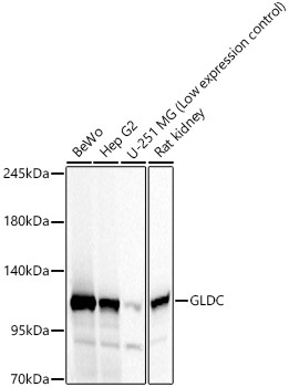
Western blot analysis of various lysates, using GLDC Rabbit pAb (CAB24541) at 1:1000 dilution. Secondary antibody: HRP-conjugated Goat anti-Rabbit IgG (H+L) (CABS014) at 1:10000 dilution. Lysates/proteins: 25μg per lane. Blocking buffer: 3% nonfat dry milk in TBST. Detection: ECL Basic Kit (AbGn00020). Exposure time: 10s.
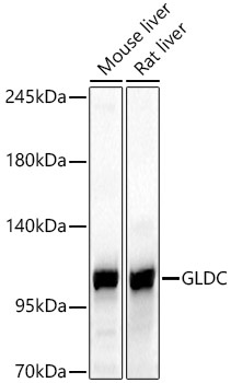
Western blot analysis of various lysates, using GLDC Rabbit pAb (CAB24541) at 1:1000 dilution. Secondary antibody: HRP-conjugated Goat anti-Rabbit IgG (H+L) (CABS014) at 1:10000 dilution. Lysates/proteins: 25μg per lane. Blocking buffer: 3% nonfat dry milk in TBST. Detection: ECL Basic Kit (AbGn00020). Exposure time: 30s.
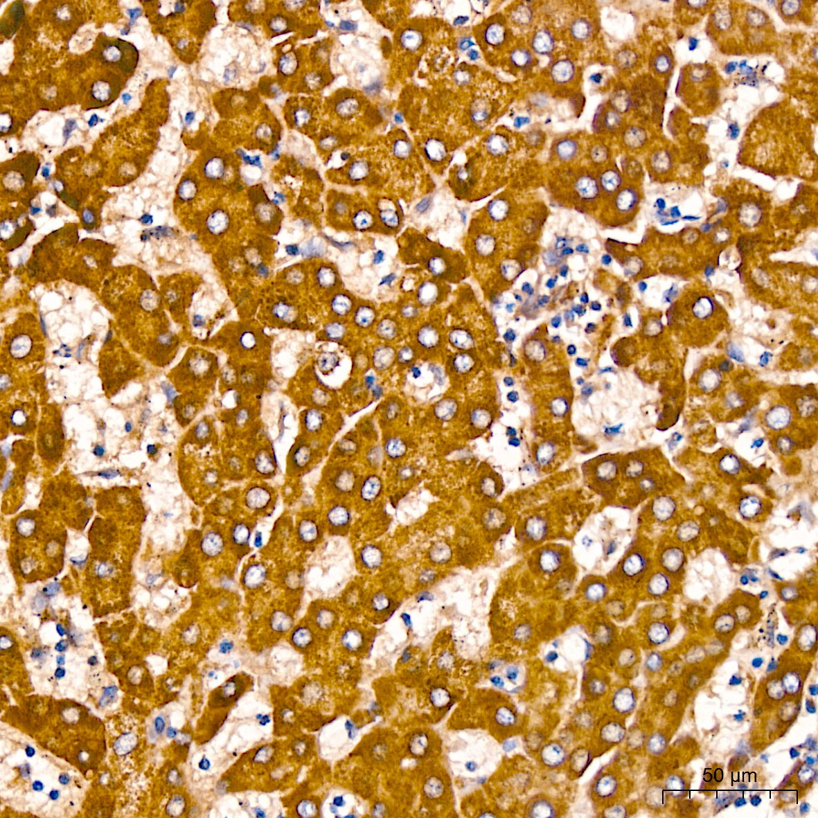
Immunohistochemistry analysis of paraffin-embedded Human liver using GLDC Rabbit pAb (CAB24541) at dilution of 1:100 (40x lens). High pressure antigen retrieval performed with 0.01M Citrate buffer (pH 6.0) prior to IHC staining.
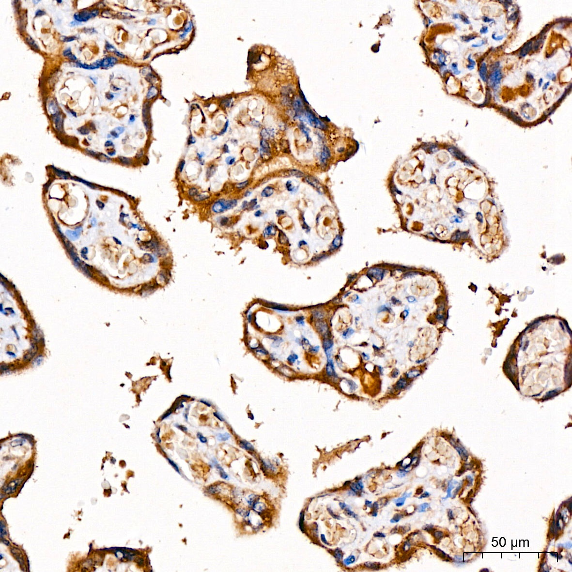
Immunohistochemistry analysis of paraffin-embedded Human placenta using GLDC Rabbit pAb (CAB24541) at dilution of 1:100 (40x lens). High pressure antigen retrieval performed with 0.01M Citrate buffer (pH 6.0) prior to IHC staining.
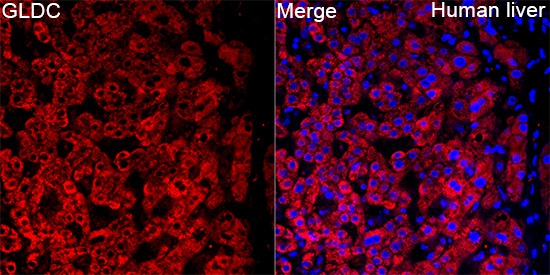
Immunofluorescence analysis of paraffin-embedded Human liver tissue using GLDC Rabbit pAb (CAB24541) at a dilution of 1:100 (40x lens). Secondary antibody: Cy3-conjugated Goat anti-Rabbit IgG (H+L) (CABS007) at 1:500 dilution. Blue: DAPI for nuclear staining. Perform high pressure antigen retrieval with 0.01 M citrate buffer (pH 6.0) prior to IF staining.
")
")


")
")