The CENPF Polyclonal Antibody (CAB18644) is an essential tool for researchers studying the centromere protein F (CENPF), a key player in cell division and chromosome segregation. This antibody, produced in rabbits, exhibits high reactivity with human samples and has been validated for Western blot applications.CENPF is a protein that localizes to the kinetochore during mitosis and is crucial for maintaining proper chromosome alignment and segregation. Its dysregulation can lead to chromosomal instability and contribute to tumorigenesis. Therefore, understanding the function and regulation of CENPF is vital for advancing our knowledge of cell division processes and identifying potential targets for cancer therapy.
With the CENPF Polyclonal Antibody, researchers can detect and analyze CENPF expression in various cell types, making it an ideal tool for studies in cell biology, cancer research, and drug development. By elucidating the role of CENPF in cellular processes, researchers can pave the way for innovative therapeutic strategies targeting this protein in cancer treatment.
Product Name:
CENPF Rabbit Polyclonal Antibody
SKU:
CAB18644
Size:
20uL, 100uL
Isotype:
IgG
Host Species:
Rabbit
Reactivity:
Human,Mouse
Immunogen:
Recombinant fusion protein containing a sequence corresponding to amino acids 1730-1890 of human CENPF (NP_057427.3).
axoneme, centrosome, ciliary basal body, ciliary transition fiber, cytoplasm, cytosol, nuclear envelope, nuclear matrix, nucleoplasm, nucleus, perinuclear region of cytoplasm, pronucleus, spindle, spindle pole
Calculated MW:
358kDa
Observed MW:
330kDa
This gene encodes a protein that associates with the centromere-kinetochore complex. The protein is a component of the nuclear matrix during the G2 phase of interphase. In late G2 the protein associates with the kinetochore and maintains this association through early anaphase. It localizes to the spindle midzone and the intracellular bridge in late anaphase and telophase, respectively, and is thought to be subsequently degraded. The localization of this protein suggests that it may play a role in chromosome segregation during mitotis. It is thought to form either a homodimer or heterodimer. Autoantibodies against this protein have been found in patients with cancer or graft versus host disease.
Purification Method:
Affinity purification
Gene ID:
1063
Storage Buffer:
Store at -20℃. Avoid freeze / thaw cycles.Buffer: PBS with 0.01% thimerosal,50% glycerol,pH7.3.
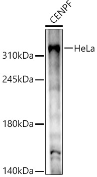
Western blot analysis of HeLa, using CENPF Rabbit pAb (CAB18644) at 1:1000 dilution.Secondary antibody: HRP Goat Anti-Rabbit IgG (H+L) (CABS014) at 1:10000 dilution.Lysates/proteins: 25μg per lane.Blocking buffer: 3% nonfat dry milk in TBST.Detection: ECL Basic Kit (AbGn00020).Exposure time: 60s.