The CAPN5 Antibody (CAB7428) is a high-quality antibody developed for reliable detection and analysis of target proteins. This antibody, produced in rabbits, is highly specific to human samples and has been validated for use in Western blot applications. By binding to calpain-5, this antibody enables accurate detection and analysis in various cell types, making it an ideal choice for studies in molecular biology and cell signaling pathways.Calpain-5, also known as calcium-dependent cysteine protease, is a key player in regulating cell growth and survival through its ability to cleave specific protein targets. Its diverse functions make it a promising target for research into diseases such as cancer, neurodegenerative disorders, and diabetes.
This antibody is validated for use in WB, IF/ICC, ELISA applications and has demonstrated reactivity against Human, Mouse, Rat samples.
Product Name:
CAPN5 Antibody
SKU:
CAB7428
Size:
20μL, 100μL
Reactivity:
Human, Mouse, Rat
Conjugate:
Unconjugated
Immunogen:
Recombinant protein (or fragment).This information is considered to be commercially sensitive.
Calpains are calcium-dependent cysteine proteases involved in signal transduction in a variety of cellular processes. A functional calpain protein consists of an invariant small subunit and 1 of a family of large subunits. CAPN5 is one of the large subunits. Unlike some of the calpains, CAPN5 and CAPN6 lack a calmodulin-like domain IV. Because of the significant similarity to Caenorhabditis elegans sex determination gene tra-3, CAPN5 is also called as HTRA3.
Purification Method
Affinity purification
Gene ID
726
RRID
AB_2767956
Buffer Information
Store at -20℃. Avoid freeze / thaw cycles. Buffer: PBS containing 50% glycerol, preserved with proclin300 or sodium azide, pH 7.3.
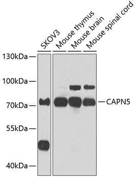
Western blot analysis of various lysates using CAPN5 Rabbit pAb (CAB7428) at 1:1000 dilution. Secondary antibody: HRP-conjugated Goat anti-Rabbit IgG (H+L) (CABS014) at 1:10000 dilution. Lysates/proteins: 25μg per lane. Blocking buffer: 3% nonfat dry milk in TBST. Detection: ECL Enhanced Kit (AbGn00021). Exposure time: 30s.
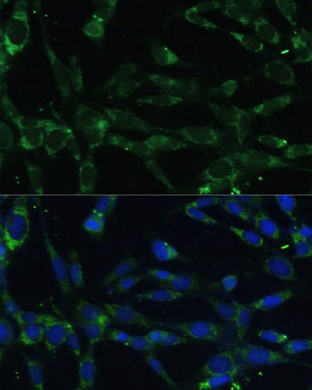
Immunofluorescence analysis of NIH-3T3 cells using CAPN5 Rabbit pAb (CAB7428) at dilution of 1:100. Secondary antibody: Cy3-conjugated Goat anti-Rabbit IgG (H+L) (CABS007) at 1:500 dilution. Blue: DAPI for nuclear staining.