The [KO Validated] N-Cadherin Antibody (CAB3045) is a high-quality antibody developed for reliable detection and analysis of target proteins. This antibody, generated in rabbits, demonstrates high specificity and sensitivity for detecting Cadherin-2 in human samples. Validated for use in Western blotting applications, it facilitates the visualization and analysis of Cadherin-2 expression in various cell types.Cadherin-2, also known as N-Cadherin, plays a pivotal role in cell-cell adhesion and signaling processes essential for tissue development and maintenance. Dysregulation of Cadherin-2 expression has been implicated in various diseases, including cancer and neurological disorders.
This antibody is validated for use in WB, IHC-P, ELISA, IF-P applications and has demonstrated reactivity against Human, Mouse, Rat samples.
Product Name:
[KO Validated] N-Cadherin Antibody
SKU:
CAB3045
Size:
20μL, 100μL
Reactivity:
Human, Mouse, Rat
Conjugate:
Unconjugated
Immunogen:
Recombinant protein (or fragment).This information is considered to be commercially sensitive.
HeLa, A-549, 293T, Mouse liver, Mouse brain, Mouse heart, Rat liver, Rat heart, Mouse heart
Cellular Localization:
Cell Membrane, Single-Pass Type I Membrane Protein.
Calculated MW:
100kDa
Observed MW:
80kDa/135kDa/130kDa
This gene encodes a classical cadherin and member of the cadherin superfamily. Alternative splicing results in multiple transcript variants, at least one of which encodes a preproprotein is proteolytically processed to generate a calcium-dependent cell adhesion molecule and glycoprotein. This protein plays a role in the establishment of left-right asymmetry, development of the nervous system and the formation of cartilage and bone.
Purification Method
Affinity purification
Gene ID
1000
RRID
AB_2863024
Buffer Information
Store at -20℃. Avoid freeze / thaw cycles. Buffer: PBS with 0.09% Sodium azide,50% glycerol,pH7.3.
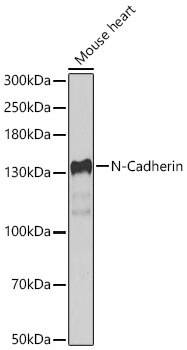
Western blot analysis of lysates from Mouse heart using N-Cadherin Rabbit pAb (CAB3045) at 1:1000 dilution. Secondary antibody: HRP-conjugated Goat anti-Rabbit IgG (H+L) (CABS014) at 1:10000 dilution. Lysates/proteins: 25 μg per lane. Blocking buffer: 3% nonfat dry milk in TBST. Detection: ECL Basic Kit (AbGn00020). Exposure time:30s.
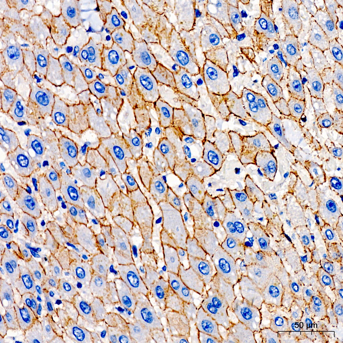
Immunohistochemistry analysis of paraffin-embedded Human liver tissue using N-Cadherin Rabbit pAb (CAB3045) at a dilution of 1:100 (40x lens). High pressure antigen retrieval was performed with 0.01 M citrate buffer (pH 6.0) prior to IHC staining.
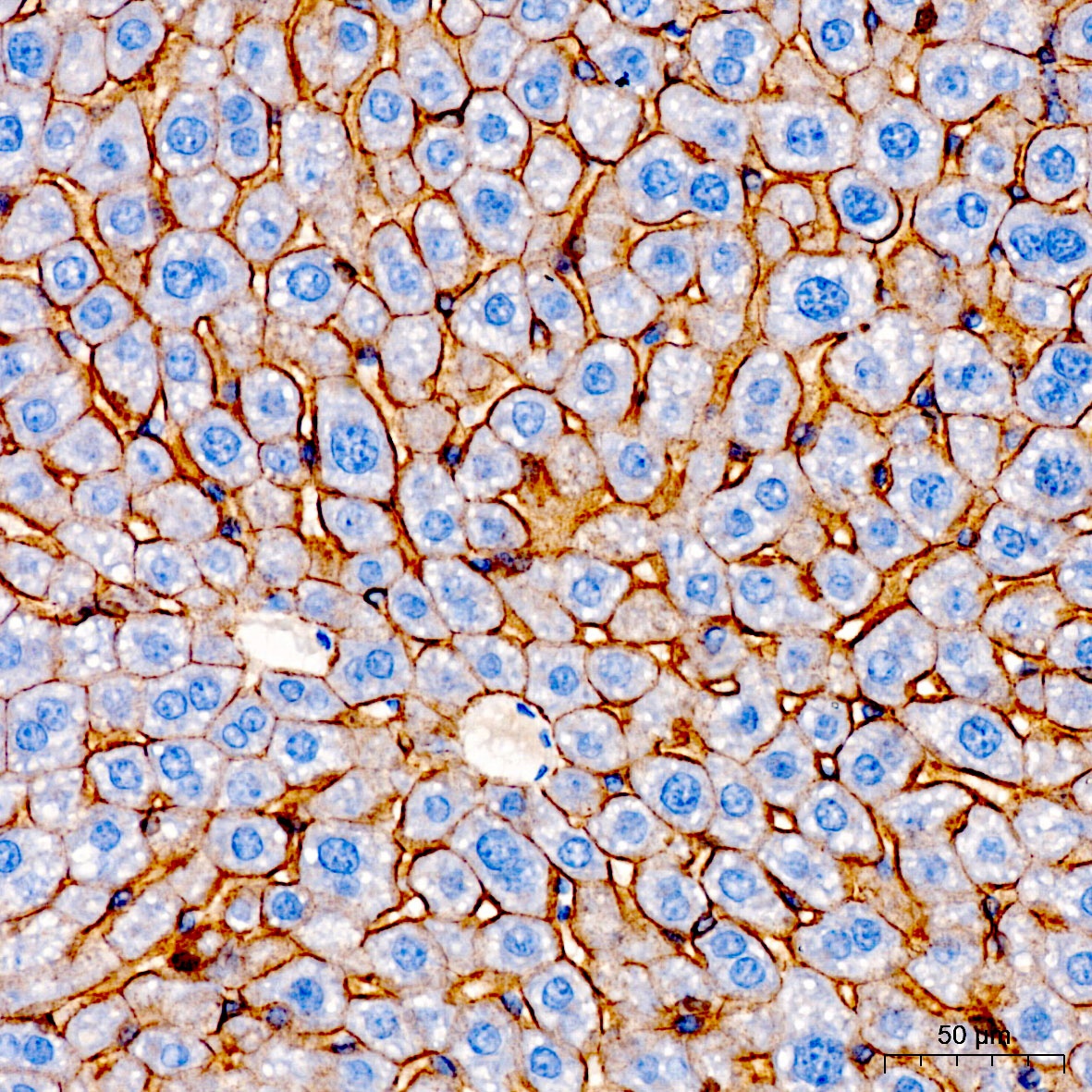
Immunohistochemistry analysis of paraffin-embedded Mouse liver tissue using N-Cadherin Rabbit pAb (CAB3045) at a dilution of 1:100 (40x lens). High pressure antigen retrieval was performed with 0.01 M citrate buffer (pH 6.0) prior to IHC staining.
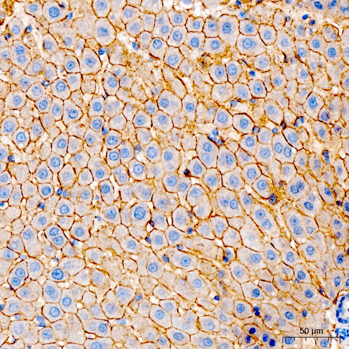
Immunohistochemistry analysis of paraffin-embedded Rat liver tissue using N-Cadherin Rabbit pAb (CAB3045) at a dilution of 1:100 (40x lens). High pressure antigen retrieval was performed with 0.01 M citrate buffer (pH 6.0) prior to IHC staining.
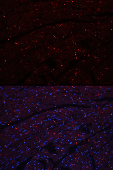
Immunofluorescence analysis of paraffin-embedded mouse heart using N-Cadherin Rabbit pAb (CAB3045). Secondary antibody: Cy3-conjugated Goat anti-Rabbit IgG (H+L) (CABS007) at 1:500 dilution. Blue: DAPI for nuclear staining.