The ATG4A Polyclonal Antibody (CAB21356) is a high-quality antibody developed for reliable detection and analysis of target proteins. This antibody, raised in rabbits, is highly specific and reacts with human samples, making it ideal for studies in autophagy, cell biology, and disease mechanisms. ATG4A is a cysteine protease that regulates autophagosome formation by cleaving the LC3/GABARAP proteins. Dysregulation of ATG4A has been implicated in various diseases, including cancer, neurodegenerative disorders, and infectious diseases. Understanding the role of ATG4A in autophagy is crucial for developing targeted therapies that aim to modulate this pathway in disease contexts.
This antibody is validated for use in WB, ELISA applications and has demonstrated reactivity against Human samples.
Product Name:
ATG4A Polyclonal Antibody
SKU:
CAB21356
Size:
20μL, 100μL
Reactivity:
Human
Conjugate:
Unconjugated
Immunogen:
Recombinant protein (or fragment).This information is considered to be commercially sensitive.
Recommended starting concentration is 1 μg/mL. Please optimize the concentration based on your specific assay requirements.
Synonyms:
APG4A, AUTL2, HsAPG4A, ATG4A
Positive Sample:
Jurkat, K-562, HeLa, Jurkat, HeLa
Cellular Localization:
Cytoplasm.
Calculated MW:
45kDa
Observed MW:
45kDa/50kDa
Autophagy is the process by which endogenous proteins and damaged organelles are destroyed intracellularly. Autophagy is postulated to be essential for cell homeostasis and cell remodeling during differentiation, metamorphosis, non-apoptotic cell death, and aging. Reduced levels of autophagy have been described in some malignant tumors, and a role for autophagy in controlling the unregulated cell growth linked to cancer has been proposed. This gene encodes a member of the autophagin protein family. The encoded protein is also designated as a member of the C-54 family of cysteine proteases.
Purification Method
Affinity purification
Gene ID
115201
Buffer Information
Store at -20℃. Avoid freeze / thaw cycles. Buffer: PBS containing 50% glycerol, preserved with proclin300 or sodium azide, pH 7.3.
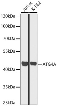
Western blot analysis of various lysates using [KO Validated] ATG4A Rabbit pAb (CAB21356) at 1:1000 dilution. Secondary antibody: HRP-conjugated Goat anti-Rabbit IgG (H+L) (CABS014) at 1:10000 dilution. Lysates / proteins: 25 μg per lane. Blocking buffer: 3 % nonfat dry milk in TBST. Detection: ECL Basic Kit (AbGn00020). Exposure time: 30s.
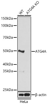
Western blot analysis of lysates from wild type (WT) and ATG4A knockout (KO) HeLa cells using [KO Validated] ATG4A Rabbit pAb (CAB21356) at 1:1000 dilution. Secondary antibody: HRP-conjugated Goat anti-Rabbit IgG (H+L) (CABS014) at 1:10000 dilution.Lysates/proteins: 25 μg per lane. Blocking buffer: 3% nonfat dry milk in TBST. Detection: ECL Basic Kit (AbGn00020). Exposure time: 5s.
")
")

![Anti-ATG4A [R05-3H3] Monoclonal Antibody (AGMB00495)](https://cdn11.bigcommerce.com/s-h68l9z2lnx/images/stencil/590x590/products/271784/693145/anti-atg4a-r05-3h3-monoclonal-antibody-agmb00495__45057.1774508373.jpg?c=2 "Anti-ATG4A [R05-3H3] Monoclonal Antibody (AGMB00495)")
![Anti-ATG4A [R07-3J5] Monoclonal Antibody (AGMB00496)](https://cdn11.bigcommerce.com/s-h68l9z2lnx/images/stencil/590x590/products/271785/691633/anti-atg4a-r07-3j5-monoclonal-antibody-agmb00496__62901.1774503578.jpg?c=2 "Anti-ATG4A [R07-3J5] Monoclonal Antibody (AGMB00496)")

")