The AKR7A2 Antibody (CAB1227) is a high-quality antibody developed for reliable detection and analysis of target proteins. This antibody, raised in rabbits, is highly specific to human samples and has been validated for use in Western blot applications. By binding to the AKR7A2 protein, this antibody enables precise detection and analysis in a variety of cell types, making it essential for studies in toxicology, pharmacology, and drug metabolism research.AKR7A2 is a member of the aldo-keto reductase superfamily and demonstrates significant activity in metabolizing various endogenous and exogenous compounds, including aldehydes and ketones.
This antibody is validated for use in WB, ELISA applications and has demonstrated reactivity against Human, Mouse samples.
Product Name:
AKR7A2 Antibody
SKU:
CAB1227
Size:
20μL, 100μL
Reactivity:
Human, Mouse
Conjugate:
Unconjugated
Immunogen:
Recombinant protein (or fragment).This information is considered to be commercially sensitive.
Recommended starting concentration is 1 μg/mL. Please optimize the concentration based on your specific assay requirements.
Synonyms:
AFAR, AKR7, AFAR1, AFB1-AR1, AKR7A2
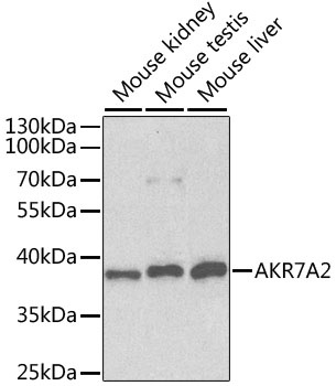
Positive Sample:
Mouse kidney, Mouse testis, Mouse liver
Cellular Localization:
Cytoplasm, Golgi Apparatus.
Calculated MW:
40kDa
Observed MW:
35kDa
The protein encoded by this gene belongs to the aldo/keto reductase (AKR) superfamily and AKR7 family, which are involved in the detoxification of aldehydes and ketones. The AKR7 family consists of 3 genes that are present in a cluster on the p arm of chromosome 1. This protein, thought to be localized in the golgi, catalyzes the NADPH-dependent reduction of succinic semialdehyde to the endogenous neuromodulator, gamma-hydroxybutyrate. It may also function as a detoxication enzyme in the reduction of aflatoxin B1 and 2-carboxybenzaldehyde. Alternative splicing results in multiple transcript variants.
Purification Method
Affinity purification
Gene ID
8574
RRID
AB_2759137
Buffer Information
Store at -20℃. Avoid freeze / thaw cycles. Buffer: PBS containing 50% glycerol, preserved with proclin300 or sodium azide, pH 7.3.
Western blot analysis of various lysates using AKR7A2 Rabbit pAb (CAB1227) at 1:1000 dilution. Secondary antibody: HRP-conjugated Goat anti-Rabbit IgG (H+L) (CABS014) at 1:10000 dilution. Lysates/proteins: 25μg per lane. Blocking buffer: 3% nonfat dry milk in TBST. Detection: ECL Basic Kit (AbGn00020).