The alpha-Smooth Muscle Actin (ACTA2) Antibody (CAB1011) is a high-quality antibody developed for reliable detection and analysis of target proteins. This antibody, generated in rabbits, exhibits high reactivity towards human samples and is validated for use in Western blot applications.ACTA2 is an essential component of smooth muscle cells, playing a crucial role in various physiological processes such as vascular contraction, organ development, and wound healing. Dysregulation of ACTA2 expression has been linked to diseases like thoracic aortic aneurysms and dissections, as well as smooth muscle cell tumors.
This antibody is validated for use in WB, IHC-P, ELISA, IF-P applications and has demonstrated reactivity against Human, Mouse, Rat samples.
Product Name:
alpha-Smooth Muscle Actin (ACTA2) Antibody
SKU:
CAB1011
Size:
20μL, 100μL
Reactivity:
Human, Mouse, Rat
Conjugate:
Unconjugated
Immunogen:
Recombinant protein (or fragment).This information is considered to be commercially sensitive.
Recommended starting concentration is 1 μg/mL. Please optimize the concentration based on your specific assay requirements.
Synonyms:
ACTSA, α-Smooth Muscle Actin (ACTA2)
Positive Sample:
Mouse heart
Cellular Localization:
Cytoplasm, Cytoskeleton.
Calculated MW:
42kDa
Observed MW:
42kDa
This gene encodes one of six different actin proteins. Actins are highly conserved proteins that are involved in cell motility, structure, integrity, and intercellular signaling. The encoded protein is a smooth muscle actin that is involved in vascular contractility and blood pressure homeostasis. Mutations in this gene cause a variety of vascular diseases, such as thoracic aortic disease, coronary artery disease, stroke, and Moyamoya disease, as well as multisystemic smooth muscle dysfunction syndrome.
Purification Method
Affinity purification
Gene ID
59
RRID
AB_2757633
Buffer Information
Store at -20℃. Avoid freeze / thaw cycles. Buffer: PBS containing 50% glycerol, preserved with proclin300 or sodium azide, pH 7.3.
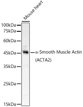
Western blot analysis of lysates from Mouse heart, using α-Smooth Muscle Actin (ACTA2) Rabbit pAb (CAB1011) at 1:800 dilution. Secondary antibody: HRP-conjugated Goat anti-Rabbit IgG (H+L) (CABS014) at 1:10000 dilution. Lysates/proteins: 25μg per lane. Blocking buffer: 3% nonfat dry milk in TBST. Detection: ECL Basic Kit (AbGn00020). Exposure time: 10s.
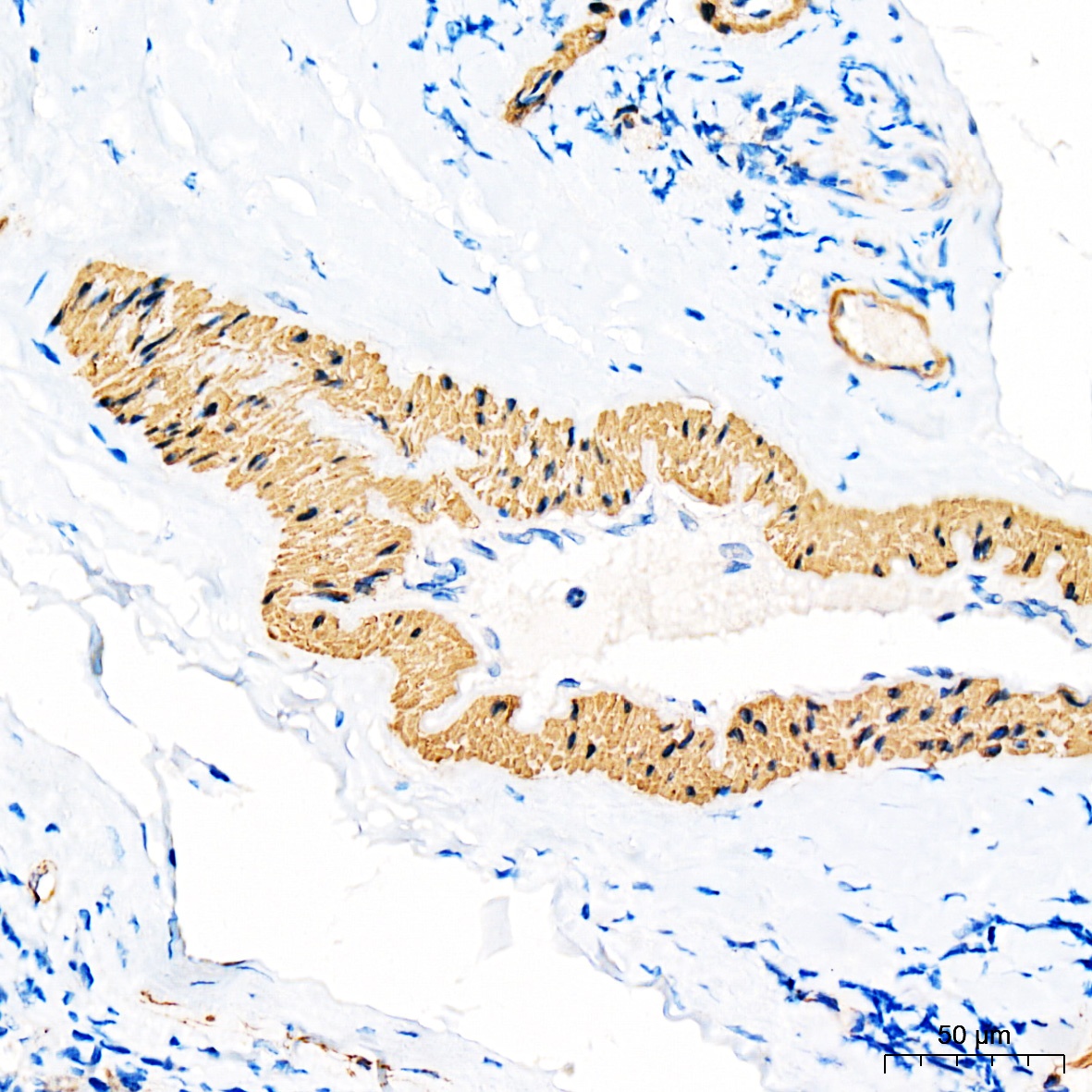
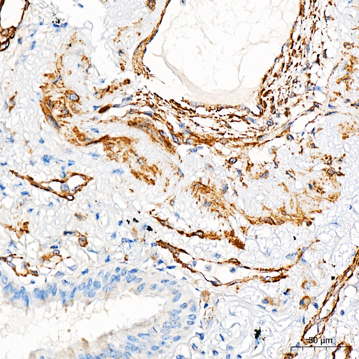
Immunohistochemistry analysis of paraffin-embedded Human tonsil tissue using α-Smooth Muscle Actin (ACTA2) Rabbit pAb (CAB1011) at a dilution of 1:1000 (40x lens). High pressure antigen retrieval was performed with 0.01 M Tris-EDTA buffer (pH 9.0) prior to IHC staining.
Immunohistochemistry analysis of paraffin-embedded Human lung tissue using α-Smooth Muscle Actin (ACTA2) Rabbit pAb (CAB1011) at a dilution of 1:1000 (40x lens). High pressure antigen retrieval was performed with 0.01 M Tris-EDTA buffer (pH 9.0) prior to IHC staining.
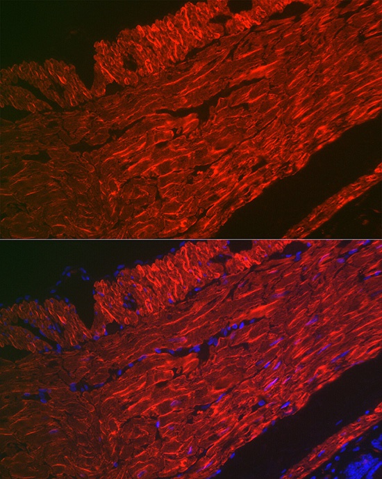
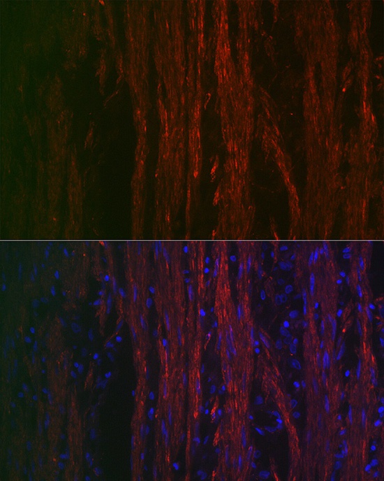
Immunofluorescence analysis of paraffin-embedded rat rectum using α-Smooth Muscle Actin (ACTA2) Rabbit pAb (CAB1011) at dilution of 1:100 (40x lens). Secondary antibody: Cy3-conjugated Goat anti-Rabbit IgG (H+L) (CABS007) at 1:500 dilution. Blue: DAPI for nuclear staining.
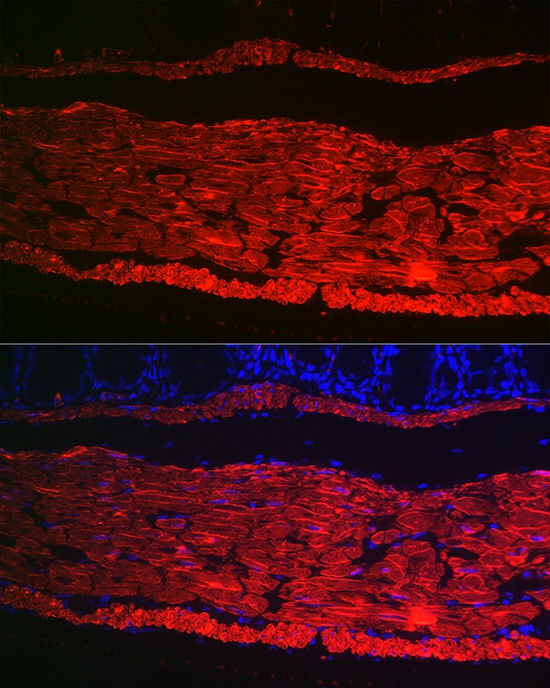
Immunofluorescence analysis of paraffin-embedded mouse colon using α-Smooth Muscle Actin (ACTA2) Rabbit pAb (CAB1011) at dilution of 1:100 (40x lens). Secondary antibody: Cy3-conjugated Goat anti-Rabbit IgG (H+L) (CABS007) at 1:500 dilution. Blue: DAPI for nuclear staining.
Immunofluorescence analysis of paraffin-embedded mouse colon using α-Smooth Muscle Actin (ACTA2) Rabbit pAb (CAB1011) at dilution of 1:100 (40x lens). Secondary antibody: Cy3-conjugated Goat anti-Rabbit IgG (H+L) (CABS007) at 1:500 dilution. Blue: DAPI for nuclear staining.


")
")
")

