
Validation of MycoGenie Rapid Mycoplasma Detection Kit - A highly sensitive visual determination method for Mycoplasma detection.
The MycoGenie Rapid Mycoplasma Detection Kit enables the detection of 28 Mycoplasma species in under 1 hour using a simple visual determination method, eliminating the need for expensive equipment and complex protocols. The kit demonstrated a detection limit of 3.07 × 10² Mycoplasma copies, matching the sensitivity of the "Gold Standard" qPCR method. The assay utilizes an isothermal amplification method with a colorimetric readout, allowing easy, rapid, and reliable detection without specialized training. The MycoGenie kit is compatible with a wide range of cell types and culture media, making it a versatile solution for cell culture laboratories. Mycoplasma contamination was shown to negatively impact cell viability and protein production, reinforcing the need for routine screening with fast and effective detection methods like MycoGenie.
Key Findings:
- The MycoGenie Rapid Mycoplasma Detection Kit enables the detection of 28 Mycoplasma species in under 1 hour using a simple visual determination method, eliminating the need for expensive equipment and complex protocols.
- The kit demonstrated a detection limit of 3.07 × 10² Mycoplasma copies, matching the sensitivity of the "Gold Standard" qPCR method.
- The assay utilizes an isothermal amplification method with a colorimetric readout, allowing easy, rapid, and reliable detection without specialized training.
- The MycoGenie kit is compatible with a wide range of cell types and culture media, making it a versatile solution for cell culture laboratories.
- Mycoplasma contamination was shown to negatively impact cell viability and protein production, reinforcing the need for routine screening with fast and effective detection methods like MycoGenie.
Validation of MycoGenie Rapid Mycoplasma Detection Kit – A highly sensitive visual determination method for Mycoplasma detection.
Introduction
Mycoplasma contamination represents one of the greatest threats to cell culture integrity resulting in severe economic less for cell culture scientists in the bio-pharmaceutical industry. Mycoplasma have deleterious effects on cultured cells as they can compete for nutrients, alter ATP levels and elicit global changes in the gene expression profile among others. Research has shown that up to 85% of cell lines can be contaminated depending on the laboratory.
Therefore, with recent medical advances from vaccine & therapeutic antibody production to CAR-T therapies depending on vibrant cell culture processes, having a Mycoplasma-free cell culture remains paramount to optimised protein production.
Currently, methods to detect Mycoplasma include labour-intensive Quantitative PCR (qPCR) and luminescent-based approaches which are unsuitable for many laboratories & raise significant challenges for cell culture scientists.
However, the recently developed MycoGenie Rapid Mycoplasma Detection Kit enables the highly sensitive detection of 28 Mycoplasma species in under 1 hour from cell culture via a simple visual determination readout thereby eliminating the need for expensive equipment and time-consuming protocols
In this study, we show that the limit of detection of MycoGenie Rapid Mycoplasma Detection kit is 3.07 x 10² copies of Mycoplasma which is similar to the “Gold Standard” qPCR method.
Mycoplasma | General Overview
The name Mycoplasma is derived from the Greek words mykes and plasma meaning "a fungus containing thread," and "a moldlike nature" and are the smallest bacteria known. They are implicated in human, animal, insect & plant diseases including AIDS, HIV and pneumoniae.
Mycoplasma contamination is a significant issue for cell culture scientists and can have a severe impact on protein production & cell health. They can be transmitted by aerosols from cultured cell lines, contaminated tissue, cultured media and biological waste such as carcasses, aborted fetal tissue and embryos that have been infected or not treated with antibiotics.
Mycoplasma contaminated cell cultures tend to be discarded which can result in a severe economic loss for the bio-pharmaceutical industry. Alternatively, scientists can use newly developed elimination reagents such as the Assay Genie MycoGenie MycoPlasma Elimination Kit that removes Mycoplasma without the need to discard precious cell lines and cultures.
Page 1
Cell Culture Contamination
Mycoplasma-infected cells produce sub-optimal amounts of protein and increase the rate at which those cells die. Therefore, Mycoplasma contamination can reduce both the quantity and quality of protein produced by cultured cells.
Mycoplasma contamination can rapidly reach very high levels & infected cell culture monolayers often resemble tumour growth.
These infected cell culture monolayers are often responsible for aerosolization of Mycoplasma and subsequent contamination of cell culture equipment, plasticware and cell lines.
Cultures of Mycoplasma are hard to maintain because they do not grow on traditional growth media such as blood or MacConkey agar. They require special nutrient media that may be used to grow Mycoplasma in vitro, but these cultures cannot be used to infect host cells unless cultured under stringent conditions that mimic the harshness of the hostile environment within which Mycoplasma grows best.
Mycoplasma can also contaminate laboratory rodents and research animals if Mycoplasma-free animals are not used. Mycoplasma can contaminate mice that have been genetically engineered to be deficient in or lack immune systems. Mycoplasma contamination can also arise if research animals are housed together with positive rodents (e.g., Mycoplastma pneumoniae, Mycoplastma suis, Mycoplastma ovis, etc.).
Assay Overview
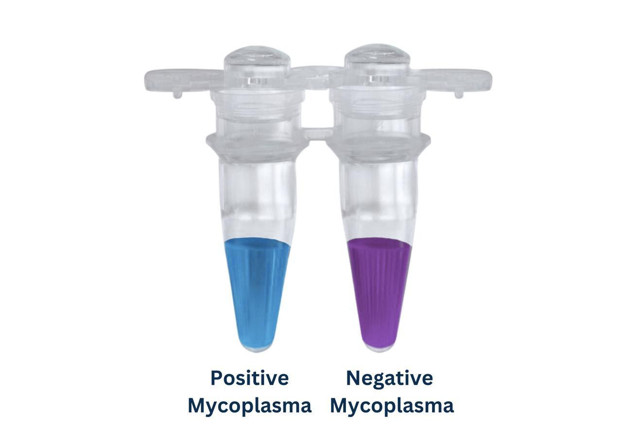
Assay Genie MycoGenie Rapid Mycoplasma Detection Kit detects Mycoplasma in cell culture using a visual determination method. The kit utilises a highly sensitive isothermal amplification method coupled with a colorimetric readout that can detect up to 28 Mycoplasma species, including 8 of the most common species associated with cell culture contamination.
Assay Features
Rapid | Detect Mycoplasma in 1 hour (28 species detected) with Isothermal amplification & pH color change that’s easily read by eye.
Simple | Easy protocol with detection by visual determination of color change meaning no expensive equipment needed.
Sensitive | Detect as little as 500cfu Mycoplasma per 1ul of cell culture supernatant.
Flexible | Detect Mycoplasma in adherent & suspensions cells such as Vero, MDCK, SP2/0, 293T, HepG2. HeLa, A549, MB-MDA231, L929, MEF, CHO, NS0, 293F, mouse hybridomas, Sf9, BHK21 & more.
Compatible | With a wide selection of cell culture media & sera such as Fetal bovine/calf serum, horse serum, Gibco KSR serum replacement & CD FortiCHO, CDM4, Expi 293 Medium, CD Hybridoma, Grace, DMEM, 1640, F12 & more.
Copy Number Determination
The performance of the MycoGenie Rapid Mycoplasma Detection to detect Mycoplasma in cell culture was extensively validated against the “Gold Standard” method of qPCR as well as conventional PCR.
In order to successfully benchmark the 3 methods, Mycoplasma copy number was first determined. Using primers specific for Mycoplasma Hyorhinis, a PCR was performed to produce a DNA standard to be used for analysis. The amplified DNA was gel extracted with a concentration of 70ng/ml.

Figure 1.
Agarose Gel showing the successful amplification of Mycoplasma Hyorhinis DNA.
Page 2
To determine the number of Mycoplasma DNA copies present in a sample, qPCR was then performed.
A dilution series was set-up using 1) gel purified Mycoplasma Hyorhinis DNA (DNA Standard) with the dilution factors of 10^-4, 10^-5, 10^-6, 10^-7, 10^-8 and 2) and a Mycoplasma stock solution (Myco Standard) at 10^-1, 10^-2 and 10^-3
Using established qPCR reagents & protocols, the ABI QuantStudio3 was used to quantify the Mycoplasma extracted DNA standard solutions and Myco standard stock solutions at the different gradients, and the standard curve was then plotted according to the quantitative experiment results and copy number.

Figure 2.
Calculation of copy number in the stock solution from the DNA standard amplification curves. Average molecular weight (MW): dsDNA = (number of base pairs) x (660 daltons/bp) = 100 bp x 660 daltons/bp = 6.6 x 10⁴ daltons = 1.98 x 10⁴ g/mol. Formula for calculating copy number: (6.02 x 10²³ copies/mol) x (concentration g/μl) x v μl / (MW g/mol) = (6.02 x 10²³ copies/mol) x (7 x 10⁻⁸ g/μl) x 1 μl / (6.6 x 10⁴ g/mol) = 6.4 x 10¹¹ copies. Note: Length of product fragment: 100 bp / Sample loading volume: 1 μl.

Table 1.
Quantitative experiment results detailing the relationship between copy number and CT value for Mycoplasma DNA Standards.

Figure 3.
Standard curve (standard), X axis: Log10 (copies), Y axis: corresponding CT value. Based on the figure, the standard curve is y = -3.569x + 42.934. R2 = 0.9982 Amplification efficiency = 10^(-1/-3.569) = 90.63% > 90%, which meets the quality requirements.
The CT values obtained from quantification of the diluted Mycoplasma stock solutions were substituted into the above standard curve equation to calculate the corresponding copy numbers, and the copy number of the Myco standard solution was then calculated according to the dilution factor.
The sample loading volume of all the diluted Myco standard solution was 1μl.
Page 3

Table 2.
Quantitative experiment results detailing the relationship between copy number and CT value for Myco standard solution.
Note 1: As the amplification curve of the Myco standard solution with a dilution factor of 10-3 overlaps with that of NTC, the amplification was deemed invalid. The CT value of the Myco standard solution with a dilution factor of 10-2 was 3.19 cycles less than that of NTC, which is in line with the “- 3” rule.
Note 2: The average copy number of the two diluted Myco standard solution above = (3.07 x 10⁵ + 4.32 x 10⁵ ) / 2 = 3.70 x 10⁵ copies, and thus, the copy number of the Myco standard solution is 3.70 x 10⁵ copies.
MycoGenie Rapid Mycoplasma Detection Validation Versus qPCR & PCR Methods
To benchmark the MycoGenie detection system against PCR as well as the “gold standard” method qPCR, a dilution series of the Myco standard stock solution was first set-up as follows: 10^-1, 10^-2, 10^-3, 10^-4 and 10^-5.
Using the diluted solutions as templates, PCR, qPCR, and MycoGenie Rapid Mycoplasma (isothermal amplification) were used to detect the Mycoplasma in each solution
Table 3.
The Myco standard stock solution dilution ratio.

PCR Detection of Mycoplasma

Figure 4.
Agarose Gel showing the detection of various copy numbers of Mycoplasma Hyorhinis DNA.
Note 1: The Myco standard solution diluted with the dilution factors of 10^-1 (3.07 x 10^4 copies) and 10^-2 (3.07 x 10^3 copies) produced obvious target amplification bands.
Note 2: There was a faint target band for the negative control, which indicates contamination of the negative control. For the Myco standard solution with dilution factors of 10^-3 (3.07 x 10^2 copies) to 10^-5 (3.07 x 10^0 copies), the band pattern was either the same as that of the negative control or there was no target band, which indicates that the solution was contaminated, and the amplification was invalid.
Page 4
QPCR Detection of Mycoplasma


Figure 5.
Calculation of copy number in the stock solution from the Mycoplasma DNA standard qPCR amplification curves.
Note 1: Amplification was noted for NTC, suggesting contamination.
Note 2: For the Myco standard stock solution with a dilution factor of 10^-3 (3.07 x 10^1 copies), the CT value on the amplification curve was 3.02 cycles less than that of NTC, which is in line with the “-3” rule. Therefore, the Mycoplasma stock solutions with dilution factors of 10^-1 (3.07 x 10^4 copies) to 10^-3 (3.07 x 10^2 copies) produced the target amplification band with smaller CT values.
Note 3: For the Myco standard stock solution with a dilution factor of 10^-4 (3.07 x 10^1 copies), the CT value on the amplification curve was close to that of NTC and differed by 0.82 cycles, suggesting contamination of the reaction solution. Therefore, the amplification was deemed invalid for Myco standard stock solution with dilution factors of 10^-4 (3.07 x 10^1 copies) and 10^-5 (3.07 x 10^0 copies).
MycoGenie Rapid Mycoplasma Detection
Visual determination of Mycoplasma copy number was performed using the MycoGenie Rapid Mycoplasma Detection kit (Isothermal PCR).

Figure 6.
Visual detection of Mycoplasma contamination using the MycoGenie Mycoplasma detection kit (Cat.no. MORV0011). ES cultured cell cultures had no detectable Mycoplasma 3 days post-treatment with the with MycoPlasma Elimination Reagent (Cat. No. MORV0012). Tube 1: Positive Control | Tube 2: Day 0 | Tube 3: Day 3 | Tube 4: Negative Control.

Table 5.
Determination of Mycoplasma copy number using the MycoGenie Rapid Detection kit (Isothermal PCR).
Note 1: In the Result interpretation column, “+” means positive, “∆” means weak positive, and “-” means negative.
Note 2: Color change was clearly noted in the Mycoplasma stock solutions with dilution factors of 10^-1 (3.07 x 10^4 copies) and 10^-2 (3.07 x 10^3 copies) in the test group.
Note 3: Slight color change was noted in the Mycoplasma stock solution with a dilution factor of 10^-3 (3.07 x 10^2 copies).
Note 4: There was virtually no color change in the Mycoplasma stock solutions with dilution factors of 10^-4 (3.07 x 10^1 copies) and 10^-5 (3.07 x 10^0 copies).
Conclusion
In conclusion, the sensitivity of the experimental protocol for qPCR is equivalent to that of isothermal amplification driven by the the MycoGenie Rapid Mycoplasma Detection kit. In detecting Mycoplasma both were able to detect Mycoplasma in stock solutions with a copy number in the hundreds. The PCR amplification protocol has lower sensitivity and was only able to detect Mycoplasma in stock solutions with a copy number in the thousands:
- The limit of detection for PCR was 3.07 x 10^3 copies of Mycoplasma.
- The limit of detection for qPCR was 3.07 x 10^2 copies of Mycoplasma.
- The limit of detection for the MycoGenie Rapid Mycoplasma Detection kit isothermal amplification was 3.07 x 10^2 copies of Mycoplasma.
Page 5
Meet Some of the Genie Team!
 Colm, CEO
 Sean, CSO
 Shane, Sales
 Beatriz, Logistics
 José, Sales
 Miren, Sales
 Natalia, Tech Support
 David, Sales
 Joe, Sales
 Marina, Tech Support
 Chris, Sales
 Shivaun, Logistics
Additional Resources


Recent Posts
-

What Are Oligodendrocytes? Functions, Markers & Disease Links
What Are Oligodendrocytes? Functions, Markers & Disease LinksOligodendrocytes are pivo …24th Sep 2025 -
Complete T Helper Cell Guide: Th1, Th2, Th17 & Functions - Your Ultimate Resource from Assay Genie
Complete T Helper Cell Guide: Th1, Th2, Th17 & Functions - Your Ultimate Resource from …27th Aug 2025 -
Apoptosis Unveiled: Your Complete Guide to Intrinsic & Extrinsic Pathways
Apoptosis Unveiled: Your Complete Guide to Intrinsic & Extrinsic PathwaysAt Assay Geni …27th Aug 2025