Understanding Radial Glial Cells: Insights into Neurodevelopmental Processes
Radial glial cells are essential in shaping the nervous system, serving dual roles in neurogenesis and structural support during the brain's developmental stages.
Key Takeaways:
- Radial glial cells are vital in neurogenesis and brain development, acting as both progenitor cells and structural guides.
- They exhibit unique radial morphology, extending from the ventricular zone to the pial surface.
- Radial glial cells differentiate into various neuronal and glial types, influencing neural circuit formation.
Table of Contents
Jump to a section:
Radial glial cells are a unique type of glial cells that play vital roles in the development and functioning of the nervous system. These remarkable cells serve as both progenitors and guides, contributing to the formation of neural circuits and providing structural support during brain development. This blog focuses on the functions, characteristics, and impacts of radial glial cells. Discover their role in neurogenesis, their interactions with other cell types, and their potential implications for brain health and disease.
Understanding Radial Glial Cells
Radial glial cells, a prominent class of glial cells found in the developing nervous system, play a fundamental role in neurogenesis and brain development. These elongated cells exhibit a unique radial morphology, extending from the ventricular zone to the pial surface. Acting as both progenitor cells and scaffolds, radial glial cells guide the migration of neurons during embryonic development. Moreover, they provide crucial structural and functional support to developing neuronal circuits. By understanding the intricate properties and functions of radial glial cells, we can resolve the intricate mechanisms underlying neural development, as well as their potential implications in various neurological conditions.
Radial Glial Cell
Types of Radial Glial Markers
| Types | Description |
| An intermediate filament protein, whose expression is upregulated during the epithelial-to-mesenchymal transition of NE cells to radial glia and persists until astrocyte development. | |
| Paired box gene 6 or paired box protein (PAX6) is a transcription factor that promotes neurogenesis. | |
| A transcription factor regulating the maintenance of radial glia. | |
| A transcription factor regulating the maintenance of radial glia. | |
| These astrocytic markers emerge as neuroepithelial cells become radial glia. | |
| Adhesion and extracellular matrix molecules: TN-C and N-cadherin | Upregulation of adhesion and extracellular matrix proteins accompanies the transformation of NE cells into radial glia. |
| An intermediate filament protein in NE cells and radial glia, whose expression persists until astrocyte development. | |
| A transcription factor and the earliest marker of the neural plate. |
Role of Glial Markers in Cell Identification
Glial markers serve as essential tools for researchers and clinicians in the field of neuroscience, enabling the identification and classification of different types of glial cells. These markers are typically proteins or molecules that exhibit selective expression within glial cell populations. By targeting these specific markers, scientists can differentiate glial cells from other cell types and gain insights into the heterogeneity and functional specialization of glia.
Radial glial cell markers, for instance, are particularly valuable in the study of radial glial cells. These markers help in visualizing the radial processes of glial cells that span across the developing neural tissue, providing a scaffold for the migration of newborn neurons. Through the identification of radial glial cell markers, researchers can trace the lineage of these cells and investigate their role in guiding neuronal migration and shaping the intricate architecture of the developing brain.
Similarly, Bergmann glia markers are employed to identify and study a specific type of glial cells known as Bergmann glia. These cells are predominantly found in the cerebellum and contribute to the organization and maintenance of the cerebellar circuitry. By utilizing Bergmann glia markers, researchers can gain insights into the functions and molecular characteristics of these specialized glial cells, elucidating their role in cerebellar development, synaptic plasticity, neuronal signaling, glutamate uptake and potassium uptake.
Immunohistochemistry, a widely used technique in neuroscience research, allows for the visualization and localization of glial markers within brain tissue samples. By coupling the specificity of glial markers with fluorescent or enzymatic labelling methods, researchers can precisely identify and map the distribution of glial cells in different brain regions. This information contributes to our understanding of the cellular composition of the nervous system and the functional roles played by glial cells in supporting neuronal function, modulating synaptic activity, and maintaining brain homeostasis.
Importance of Radial Glial Stem Cells
Radial glial stem cells represent a critical population of progenitor cells in the developing nervous system. These remarkable cells serve as a source of neural diversity and are responsible for generating a wide range of neuronal and glial cell types. During embryonic development, radial glial stem cells exhibit a dual role. Firstly, they self-renew, maintaining their own population. Secondly, they give rise to intermediate progenitor cells and ultimately differentiate into neurons or glial cells. This process, known as neurogenesis, is essential for the establishment of the complex neural circuitry that underlies brain function. Furthermore, radial glial stem cells contribute to the formation of specialized brain structures and play a key role in the development of brain regions responsible for higher cognitive functions. Understanding the importance of radial glial stem cells provides insights into the mechanisms of neural development, regeneration, and potential therapeutic strategies targeting neurodevelopmental disorders or brain injuries.
Exploring Glial Cell Development
Early neuroepithelial cells divide symmetrically to produce new neuroepithelial cells. Early neurons are probably produced by some neuroepithelial cells. Neuroepithelial cells elongate and change into radial glial (RG) cells when the growing brain epithelium thickens. The asymmetric division of RG results in the direct or indirect production of neurons via intermediate progenitor cells (nIPCs). The intermediate progenitor cells (oIPCs) that produce oligodendrocytes are another source of oligodendrocytes from RG. The brain thickness increases as the offspring of RG and IPCs go into the mantel for differentiation, lengthening RG cells even further. The apical-basal polarity of radial glia causes them to touch the ventricle apically (down), where they project a single primary cilium, and the meninges, basal lamina, and blood vessels basally (up). oIPC production continues as the majority of RG start to separate from the apical side and transform into astrocytes during the conclusion of embryonic development. A portion of RG maintain apical contact and carry on their NSC roles in the newborn. Through nIPCs and oIPCS, these neonatal RG continue to produce neurons and oligodendrocytes; some of them differentiate into ependymal cells, while others become adult SVZ astrocytes (type B cells), which continue to serve as NSCs in adults. B cells maintain an epithelial architecture with basal ends in blood arteries and apical contact at the ventricle. Through (n and o) IPCs, B cells continue to produce neurons and oligodendrocytes.
Glial nature of neural stem cells (NSCs) in development and in the adult
Related Products
Radial Glial Markers ELISA Kits
Vimentin is an intermediate filament protein that is highly expressed in radial glial cells. It is often used as a marker to identify and label radial glial cells in tissue sections or cultures. Vimentin helps visualize the characteristic radial processes of these cells and distinguish them from other cell types.

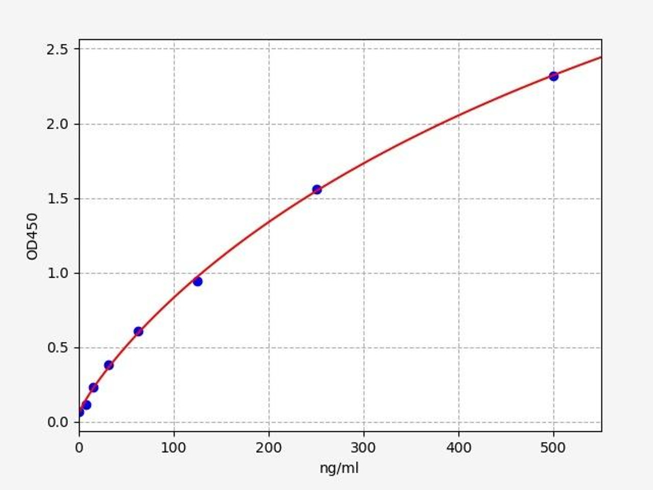
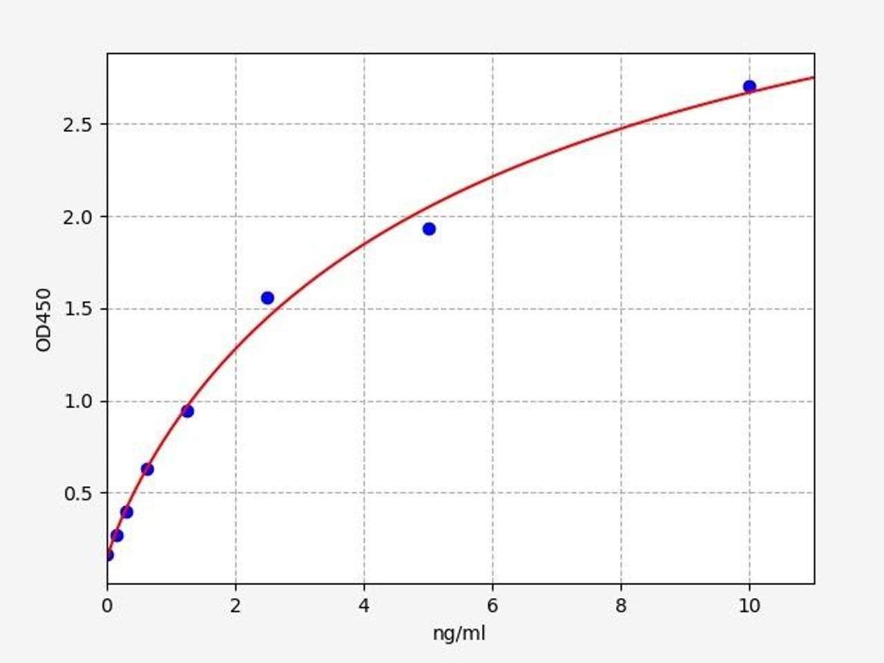
| Human Paired Box Gene 6 (PAX6) ELISA Kit | |
|---|---|
| Sensitivity | 0.144ng/mL |
| Range | 0.312-20ng/mL |
| Reactivity | Human |
Pax6 is a transcription factor involved in the development of the central nervous system, including radial glial cells. It is utilized as a marker to identify and study radial glial cells during different stages of embryonic and postnatal neurogenesis. Pax6 expression provides insights into the specification and maturation of radial glial cells.
SOX2 (SRY-Box Transcription Factor 2) protein belongs to the SOX (SRY-Box) family of transcription factors. SOX2 is a key regulator of embryonic development, involved in the formation of many tissues and organs. SOX2 is expressed in many different tissues during embryonic development, including the brain, heart, and spinal cord. Diseases associated with SOX2 include Microphthalmia, Syndromic 3 and Septooptic Dysplasia.
Functions and Impacts of Radial Glial cells
The primary function attributed to radial glial cells (RGLCs) is homeostatic neurogenesis, which refers to the generation of new neurons through precursor cells derived from RGLC proliferation and maturation. While neurogenesis is primarily associated with embryonic development, certain animals exhibit ongoing neurogenic processes throughout their lifespan. An example of adult neurogenesis was observed in the central nervous system of canaries during the breeding season. In adult rodents, RGLCs in the subgranular zone (SGZ) have been found to produce excitatory neurons associated with memory formation and learning. Similarly, RGLCs in the subventricular zone (SVZ) contribute to adult neurogenesis by generating inhibitory neurons involved in odor discrimination and odor-reward association. It is noteworthy that most of these cells are usually in a dormant or quiescent state, despite their proliferative potential. Understanding the mechanisms of quiescence and proliferation in RGLCs has become a crucial area of study. Maintaining a quiescent state is believed to preserve stemness and neurogenic potential by preventing DNA, protein, or mitochondrial damage that could lead to premature cell senescence and the loss of the stem cell niche. Researchers have focused on identifying local niche signals, such as the Notch pathway or ASCL1, that regulate quiescence. Additionally, investigations are ongoing to determine the signaling pathways responsible for guiding the differentiation of newly generated cells into neuronal or glial phenotypes.
In conclusion, radial glial cells are integral players in the intricate landscape of the nervous system. Their dual roles as progenitors and guides contribute to the development and functioning of the brain. Through neurogenesis, radial glial cells give rise to a diverse array of neuronal and glial cell types, shaping the complex neural circuitry. Their interactions with other cell types provide crucial structural support and help orchestrate the precise wiring of the brain. Moreover, research on radial glial cells continues to uncover their significance in various aspects of neural development, synaptic plasticity, and brain function. By unraveling the secrets of radial glial cells, we gain deeper insights into the fundamental processes that underlie the functioning of our remarkable nervous system. Further exploration of radial glial cells promises to unveil new avenues for understanding and potentially treating neurological disorders
Written by Pragna Krishnapur
Pragna Krishnapur completed her bachelor degree in Biotechnology Engineering in Visvesvaraya Technological University before completing her masters in Biotechnology at University College Dublin.
Recent Posts
-
Enavatuzumab: Revolutionizing Cancer Research Through Novel Therapeutics
Quick Facts About EnavatuzumabWhat is Enavatuzumab?Enavatuzumab is a monoclonal antibo …17th Dec 2025 -
Alemtuzumab: Mechanism, Applications, and Biosimilar Advancements
Quick Facts About AlemtuzumabWhat is Alemtuzumab?Alemtuzumab is a monoclonal antibody …17th Dec 2025 -
Validation of MycoGenie Rapid Mycoplasma Detection Kit - A highly sensitive visual determination method for Mycoplasma detection.
The MycoGenie Rapid Mycoplasma Detection Kit enables the detection of 28 Mycoplasma sp …3rd Mar 2025


