Oligodendrocytes: What Are They?
Oligodendrocytes are pivotal CNS cells, integral for myelin formation and nerve function, with implications in various neurological disorders.
Key Takeaways
- Oligodendrocytes are specialized glial cells in the central nervous system (CNS), crucial for myelinating axons, enhancing nerve signal transmission, and supporting neuronal function.
- They differ from Schwann cells, which myelinate in the peripheral nervous system.
- Oligodendrocytes are implicated in diseases like multiple sclerosis and Alzheimer's, with treatment strategies focusing on immunomodulation, remyelination, neuroprotection, and symptomatic relief.
What Are Oligodendrocytes?
Oligodendrocytes are a specific type of glial cell found in the central nervous system (CNS) of vertebrates, including humans. They are primarily found in the brain and spinal cord, which are the major components of the CNS. Within these regions, oligodendrocytes are distributed throughout the gray and white matter. The gray matter consists of neuronal cell bodies and unmyelinated axons, while the white matter contains myelinated axons. Oligodendrocytes are particularly abundant in the white matter, where they play a crucial role in myelinating multiple axons to facilitate efficient signal transmission. Within the CNS, oligodendrocytes are widely distributed and interact with a multitude of neurons. These cells are characterized by their numerous, branching processes that extend from their cell bodies. These processes enable oligodendrocytes to establish close contact with axons, the long, slender projections of neurons responsible for transmitting electrical signals.
Schematic of an Oligodendrocyte
What is the Function of Oligodendrocytes?
The primary function of oligodendrocytes is the production and maintenance of myelin, a specialized substance that forms an insulating sheath around axons. Myelin consists of layers of lipid-rich membranes produced by oligodendrocytes. This myelin sheath acts as an electrical insulator, significantly enhancing the efficiency and speed of nerve impulse conduction. Oligodendrocytes achieve myelination by extending their processes and wrapping them around segments of axons. Each oligodendrocyte can myelinate multiple axons, forming compact bundles of myelinated fibers. The myelin sheath created by oligodendrocytes not only prevents electrical signal leakage but also facilitates the saltatory conduction of nerve impulses. Saltatory conduction refers to the rapid "jumping" of electrical signals between nodes of Ranvier, which are small gaps in the myelin sheath along the axon. This mode of conduction allows for faster propagation of signals along the axon, conserving energy and optimizing neuronal communication.
Myelination: Oligodendrocyte Differentiation and Ensheathment of Axons
Oligodendrocyte-Neuron Interactions
Oligodendrocytes and neurons maintain a dynamic relationship crucial for proper nervous system function. This interplay involves trophic support, where oligodendrocytes provide growth factors and nutrients that promote neuronal survival and circuit formation. Additionally, oligodendrocytes play a role in energy metabolism by supplying neurons with lactate, a key metabolic substrate. This metabolic coupling ensures the energetic demands of myelinated axons and supports neuronal activity. Furthermore, oligodendrocytes contribute to synaptic plasticity, modulating the formation, maintenance, and plasticity of synapses. They influence the electrical properties of axons and release signaling molecules that shape neural circuitry, thus impacting information processing in the central nervous system (CNS). Disruptions in oligodendrocyte-neuron interactions have implications for neurological disorders. Defects in trophic support, energy metabolism, or synaptic plasticity can contribute to the pathogenesis of conditions such as multiple sclerosis and Alzheimer's disease.
Oligodendrocytes Vs Schwann Cells
Oligodendrocytes and Schwann cells are both glial cells involved in myelination, but they exhibit important differences in terms of location, myelination patterns, and developmental origins. Oligodendrocytes are found exclusively in the central nervous system, which comprises the brain and spinal cord. In contrast, Schwann cells are located in the peripheral nervous system, which includes the nerves outside of the CNS.
One significant distinction between oligodendrocytes and Schwann cells lies in their myelination patterns. Oligodendrocytes extend their processes to wrap around different segments of multiple axons, forming multiple layers of myelin sheath. This allows one oligodendrocyte to myelinate several axons simultaneously. On the other hand, Schwann cells envelop a single axon by spiraling around it, forming a single layer of myelin.
Additionally, oligodendrocytes and Schwann cells have different developmental origins. Oligodendrocytes originate from precursor cells within the CNS during embryonic development, while Schwann cells are derived from neural crest cells in the PNS. Despite these distinctions, both oligodendrocytes and Schwann cells play vital roles in supporting and insulating neurons. Their myelination processes contribute to the efficient and rapid transmission of electrical signals, ensuring the proper functioning of the nervous system.
Oligodendrocytes Related Kits

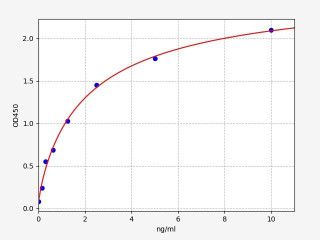
| Human OLIG2 (Oligodendrocyte transcription factor 2) ELISA Kit (HUFI07413) | |
|---|---|
| ELISA Type | Sandwich |
| Sensitivity | 0.094ng/ml |
| Range | 0.156-10ng/ml |

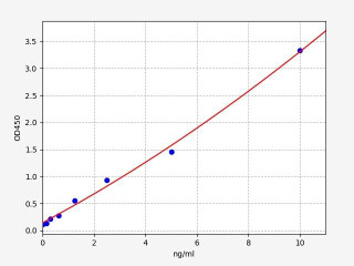
| Human MBP(myelin basic protein) ELISA Kit | |
|---|---|
| ELISA Type | Sandwich |
| Sensitivity | 0.094ng/ml |
| Range | 0.156-10ng/ml |

| Human MOG / Myelin oligodendrocyte glycoprotein ELISA Kit | |
|---|---|
| ELISA Type | Sandwich |
| Sensitivity | 0.094ng/ml |
| Range | 0.156-10ng/ml |
Oligodendrocytes Lineage Markers and Histology Techniques
Oligodendrocyte lineage markers and histology techniques play a crucial role in identifying and studying oligodendrocytes. Several marker proteins are used to pinpoint oligodendrocyte lineage cells, including:
- Oligodendrocyte transcription factor (OLIG) family proteins: These transcription factors, such as OLIG1 and OLIG2, are vital for oligodendrocyte development and function.
- Myelin basic protein (MBP): MBP is an essential protein for the formation and maintenance of the myelin sheath, a characteristic feature of oligodendrocytes.
- Oligodendrocyte-specific proteolipid protein (PLP): PLP is found in the myelin sheath and helps maintain its structural integrity.
Histology techniques are used to visualize oligodendrocytes and their characteristics. Hematoxylin and eosin (H&E) staining is commonly employed to observe the cellular morphology of oligodendrocytes. Immunohistochemistry enables the detection of specific oligodendrocyte lineage markers, including Olig-17 (a marker for oligodendrocyte progenitor cells), and CNPase (a marker for myelin-forming oligodendrocytes). Additionally, fluorescence microscopy can be utilized to visualize oligodendrocytes labeled with fluorescent probes, providing enhanced visualization capabilities.
Diseases associated with dysfunctional Oligodendrocytes
Dysfunctional oligodendrocytes are associated with various diseases, including demyelinating diseases, neurodegenerative disorders, oligodendrogliomas, and leukodystrophies. Demyelinating diseases are characterized by the breakdown of the myelin sheath, leading to impaired nerve function and development. One example is multiple sclerosis (MS), an autoimmune disorder that attacks the myelin sheath, causing demyelination and axonal damage. Cerebral palsy, resulting from brain damage during development, can also affect oligodendrocytes and lead to motor function and coordination problems. Pelizaeus-Merzbacher disease, a hereditary disorder, results in the loss of myelin-forming oligodendrocytes. Traumatic brain injury (TBI) is another condition that can damage oligodendrocytes and cause demyelination, contributing to disability.
Neurodegenerative disorders, such as Alzheimer's disease, Parkinson's disease, and Huntington's disease, involve the dysfunction of oligodendrocytes and subsequent loss of neuronal function. These diseases are characterized by problems with memory, cognition, behavior, coordination, and movement. Oligodendrogliomas are tumors derived from oligodendrocytes. Although they are usually slow-growing and rarely metastasize, they can be challenging to treat due to their aggressive nature. Leukodystrophies encompass a group of hereditary disorders affecting the myelin sheath. These conditions can lead to movement difficulties, cognitive impairment, and other neurological dysfunctions. Examples of leukodystrophies include adrenoleukodystrophy (ALD) and metachromatic leukodystrophy (MLD).
Treatment Strategies for Oligodendrocyte Disorders
Treatment strategies for oligodendrocyte disorders aim to manage symptoms and enhance the quality of life, although a cure is currently unavailable for most of these conditions. While the treatment options for oligodendrocyte disorders are still evolving, several strategies are being explored. Here are some of the treatment approaches currently considered:
- Immunomodulatory Therapies: In demyelinating disorders such as multiple sclerosis (MS), the immune system mistakenly attacks the myelin sheath. Medications known as disease-modifying therapies (DMTs) are used to modulate the immune response and reduce inflammation in order to slow disease progression and manage symptoms. Examples of DMTs include interferon-beta, glatiramer acetate, and monoclonal antibodies like natalizumab or ocrelizumab.
- Remyelination Therapies: Stimulating the regeneration of myelin is a key focus of research. Various approaches are being investigated, including the use of stem cells, which have the potential to differentiate into oligodendrocytes and promote remyelination. Other strategies involve the use of small molecules or biologics that enhance oligodendrocyte precursor cell differentiation and myelination.
- Neuroprotective Approaches: Oligodendrocyte disorders often involve neuronal damage in addition to demyelination. Protecting and preserving nerve cells from further injury is an important therapeutic goal. Neuroprotective strategies may involve the use of antioxidants, anti-inflammatory agents, or drugs that promote cell survival and repair.
- Symptomatic Treatment: Many oligodendrocyte disorders manifest with neurological symptoms that can be managed with symptomatic treatment. For example, medications may be prescribed to alleviate pain, muscle spasms, fatigue, or bladder dysfunction commonly seen in MS.
- Physical and Occupational Therapy: Rehabilitation approaches can play a significant role in optimizing function and quality of life for individuals with oligodendrocyte disorders. Physical and occupational therapies may focus on improving mobility, coordination, strength, and activities of daily living.
These treatment strategies may vary depending on the specific oligodendrocyte disorder and individual patient characteristics. The field of oligodendrocyte research is rapidly evolving, and new therapeutic strategies are continuously being explored in preclinical and clinical studies.
Written by Lauryn McLoughlin
Lauryn McLoughlin completed her undergraduate degree in Neuroscience before completing her masters in Biotechnology at University College Dublin.
Recent Posts
-
Enavatuzumab: Revolutionizing Cancer Research Through Novel Therapeutics
Quick Facts About EnavatuzumabWhat is Enavatuzumab?Enavatuzumab is a monoclonal antibo …17th Dec 2025 -
Alemtuzumab: Mechanism, Applications, and Biosimilar Advancements
Quick Facts About AlemtuzumabWhat is Alemtuzumab?Alemtuzumab is a monoclonal antibody …17th Dec 2025 -
Validation of MycoGenie Rapid Mycoplasma Detection Kit - A highly sensitive visual determination method for Mycoplasma detection.
The MycoGenie Rapid Mycoplasma Detection Kit enables the detection of 28 Mycoplasma sp …3rd Mar 2025


