The WDR36 Polyclonal Antibody (PAC020878) is a vital tool for research involving the WDR36 protein, a key player in ocular-related diseases such as glaucoma. This antibody, produced in rabbits, exhibits high reactivity towards human samples and has been verified for use in Western blotting applications. By binding specifically to the WDR36 protein, this antibody enables precise detection and analysis in a variety of cell types, making it an indispensable asset for studies in ophthalmology and vision-related research.WDR36, also known as WD repeat-containing protein 36, is essential for maintaining normal intraocular pressure and visual function.
Dysregulation of WDR36 has been linked to the development of glaucoma, a leading cause of irreversible blindness worldwide. Investigating the role of WDR36 can provide crucial insights into the pathogenesis of glaucoma and aid in the development of novel therapeutic strategies for this sight-threatening condition.By utilizing the WDR36 Polyclonal Antibody in your research, you can delve deeper into the mechanisms underlying glaucoma and other ocular disorders, potentially paving the way for improved diagnostic tools and treatment options in the field of ophthalmology.
Antibody Name:
WDR36 Antibody (PACO20878)
Antibody SKU:
PACO20878
Size:
50ul
Host Species:
Rabbit
Tested Applications:
ELISA, IHC
Recommended Dilutions:
ELISA:1:2000-1:5000, IHC:1:20-1:100
Species Reactivity:
Human
Immunogen:
Synthetic peptide of human WDR36
Form:
Liquid
Storage Buffer:
-20°C, pH7.4 PBS, 0.05% NaN3, 40% Glycerol
Purification Method:
Antigen affinity purification
Clonality:
Polyclonal
Isotype:
IgG
Conjugate:
Non-conjugated
The image on the left is immunohistochemistry of paraffin-embedded Human brain tissue using PACO20878(WDR36 Antibody) at dilution 1/20, on the right is treated with synthetic peptide. (Original magnification: x200).
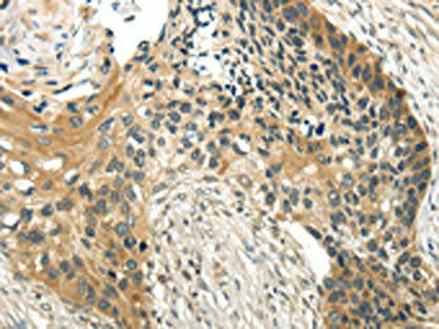
The image on the left is immunohistochemistry of paraffin-embedded Human esophagus cancer tissue using PACO20878(WDR36 Antibody) at dilution 1/20, on the right is treated with synthetic peptide. (Original magnification: x200).
Background:
This gene encodes a member of the WD repeat protein family. WD repeats are minimally conserved regions of approximately 40 amino acid, typically bracketed by gly-his and trp-asp (GH-WD), which may facilitate formation of heterotrimeric or multiprotein complexes. Members of this family are involved in a variety of cellular processes, including cell cycle progression, signal transduction, apoptosis, and gene regulation. Mutations in this gene have been associated with adult-onset primary open-angle glaucoma (POAG).
Synonyms:
WD repeat domain 36
UniProt Protein Function:
WDR36: Involved in T-cell activation and highly co-regulated with IL2. Defects in WDR36 are the cause of primary open angle glaucoma type 1G (GLC1G). Primary open angle glaucoma (POAG) is characterized by a specific pattern of optic nerve and visual field defects. The angle of the anterior chamber of the eye is open, and usually the intraocular pressure is increased. The disease is asymptomatic until the late stages, by which time significant and irreversible optic nerve damage has already taken place.
UniProt Protein Details:
Protein type:Nucleolus
Chromosomal Location of Human Ortholog: 5q22.1
Cellular Component: small subunit processome; nucleolus
Biological Process: retinal homeostasis; visual perception; response to stimulus; regulation of axon extension; rRNA processing
Disease: Glaucoma 1, Open Angle, G
NCBI Summary:
This gene encodes a member of the WD repeat protein family. WD repeats are minimally conserved regions of approximately 40 amino acids typically bracketed by gly-his and trp-asp (GH-WD), which may facilitate formation of heterotrimeric or multiprotein complexes. Members of this family are involved in a variety of cellular processes, including cell cycle progression, signal transduction, apoptosis, and gene regulation. Mutations in this gene have been associated with adult-onset primary open-angle glaucoma (POAG). [provided by RefSeq, Jul 2008]


.")


. Section was blocked with 10% normal goat serum 30min at RT. Then primary antibody (1% BSA) was incubated at 4°C overnight. The primary is detected by a Goat anti-rabbit polymer IgG labeled by HRP and visualized using 0.05% DAB.")
. Section was blocked with 10% normal goat serum 30min at RT. Then primary antibody (1% BSA) was incubated at 4°C overnight. The primary is detected by a Goat anti-rabbit polymer IgG labeled by HRP and visualized using 0.05% DAB.")
