The WBP11 Antibody (CAB16191) is a high-quality antibody developed for reliable detection and analysis of target proteins. This antibody, produced in rabbits, exhibits high reactivity with human samples and has been validated for use in Western blot applications.By targeting the WBP11 protein, this antibody enables the detection and analysis of WBP11 expression in various cell types, making it a valuable asset for studies in molecular biology and gene regulation. Understanding the function and regulation of WBP11 is crucial for advancing our knowledge of how cells control gene expression and maintain proper cellular function.
This antibody is validated for use in WB, IHC-P, IF/ICC, ELISA applications and has demonstrated reactivity against Human, Mouse, Rat samples.
Product Name:
WBP11 Antibody
SKU:
CAB16191
Size:
20μL, 100μL
Reactivity:
Human, Mouse, Rat
Conjugate:
Unconjugated
Immunogen:
Recombinant protein (or fragment).This information is considered to be commercially sensitive.
This gene encodes a nuclear protein, which colocalizes with mRNA splicing factors and intermediate filament-containing perinuclear networks. This protein has 95% amino acid sequence identity to the mouse Wbp11 protein. It contains two proline-rich regions that bind to the WW domain of Npw38, a nuclear protein, and thus this protein is also called Npw38-binding protein NpwBP. The Npw38-NpwBP complex may function as a component of an mRNA factory in the nucleus.
Purification Method
Affinity purification
Gene ID
51729
RRID
AB_2763643
Buffer Information
Store at -20℃. Avoid freeze / thaw cycles. Buffer: PBS containing 50% glycerol, preserved with proclin300 or sodium azide, pH 7.3.
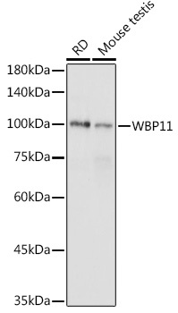
Western blot analysis of various lysates using WBP11 Rabbit pAb (CAB16191) at 1:1000 dilution. Secondary antibody: HRP-conjugated Goat anti-Rabbit IgG (H+L) (CABS014) at 1:10000 dilution. Lysates/proteins: 25μg per lane. Blocking buffer: 3% nonfat dry milk in TBST. Detection: ECL Basic Kit (AbGn00020). Exposure time: 1s.
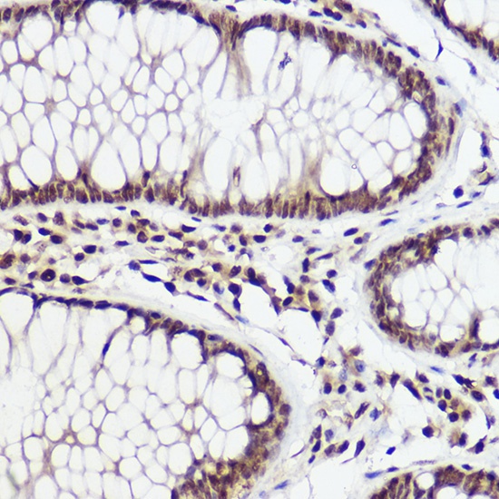
Immunohistochemistry analysis of paraffin-embedded Human colon carcinoma using WBP11 Rabbit pAb (CAB16191) at dilution of 1:100 (40x lens). High pressure antigen retrieval performed with 0.01M Citrate buffer (pH 6.0) prior to IHC staining.
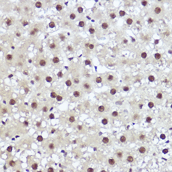
Immunohistochemistry analysis of paraffin-embedded Rat liver using WBP11 Rabbit pAb (CAB16191) at dilution of 1:100 (40x lens). High pressure antigen retrieval performed with 0.01M Citrate buffer (pH 6.0) prior to IHC staining.
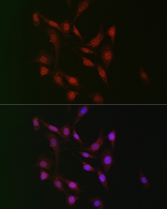
Immunofluorescence analysis of NIH/3T3 cells using WBP11 Rabbit pAb (CAB16191) at dilution of 1:100 (40x lens). Secondary antibody: Cy3-conjugated Goat anti-Rabbit IgG (H+L) (CABS007) at 1:500 dilution. Blue: DAPI for nuclear staining.