Description
VERO Host Cell Protein (HCP) ELISA Kit
The Vero HCP (Host Cell Protein) ELISA Kit is specifically developed for precise quantification of host cell protein levels in Vero cell culture samples. This kit offers exceptional sensitivity and accuracy, ensuring consistent and dependable results for various research purposes.Host cell proteins are proteins produced by the host cells used in biopharmaceutical manufacturing processes. Monitoring and quantifying these proteins are crucial for assessing the purity and safety of biotherapeutics.
The Vero HCP ELISA Kit provides a reliable tool for researchers and scientists to analyze and optimize cell culture conditions, as well as monitor protein contaminants throughout the production process. With its high performance and ease of use, the Vero HCP ELISA Kit is an indispensable tool for biopharmaceutical research and development, contributing to the advancement of bioprocessing technologies and the production of safe and effective biotherapeutics.
| Product Name: | VERO Host Cell Protein (HCP) ELISA Kit |
| Product Code: | AEFI00494 |
| Size: | 96T |
| Alias: | Vero HCPs, VERO host cell proteins |
| Detection method: | Sandwich ELISA, Double Antibody |
| Application: | VERO HCPs ELISA Kit allows for the in vitro quantitative determination of Vero HCPs concentrations in serum, plasma, tissue homogenates and other biological fluids. |
| Sensitivity: | < 0.188ng/ml |
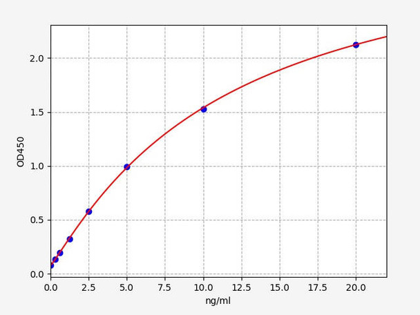
| Range: | 0.313-20ng/ml |
| Storage: | 2-8°C for 6 months |
| Note: | For Research Use Only |
This kit was based on sandwich enzyme-linked immune-sorbent assay technology. Anti Vero HCPs antibody was pre-coated onto the 96-well plate. The biotin conjugated anti Vero HCPs antibody was used as the detection antibody. The standards and pilot samples were added to the wells subsequently. After incubation, unbound conjugates were removed by wash buffer. Then, biotinylated detection antibody was added to bind with Vero HCPs conjugated on coated antibody. After washing off unbound conjugates, HRP-Streptavidin was added. After a third washing, TMB substrates were added to visualize HRP enzymatic reaction. TMB was catalyzed by HRP to produce a blue color product that turned yellow after adding a stop solution. Read the O.D. absorbance at 450nm in a microplate reader. The concentration of Vero HCPs in the sample was calculated by drawing a standard curve. The concentration of the target substance is proportional to the OD450 value.
| Recovery: | Matrices listed below were spiked with certain level of Vero HCPs and the recovery rates were calculated by comparing the measured value to the expected amount of Vero HCPs in samples. | ||||||
| |||||||
| CV(%): | Intra-Assay: CV<8% Inter-Assay: CV<10% |
When carrying out an ELISA assay it is important to prepare your samples in order to achieve the best possible results. Below we have a list of procedures for the preparation of samples for different sample types.
| Sample Type | Protocol |
| Tissue Sample | Generally tissue samples are required to be made into homogenization. Protocol is as below: 1.1. Place the target tissue on the ice. Remove residual blood by washing tissue with pre-cooling PBS buffer (0.01M, pH=7.4). Then weigh for usage. 1.2. Use lysate to grind tissue homogenates on the ice. The adding volume of lysate depends on the weight of the tissue. Usually, 9mL PBS would be appropriate to 1 gram tissue pieces. Some protease inhibitors are recommended to add into the PBS (e.g. 1mM PMSF). 1.3. Do further process using ultrasonic disruption or freeze-thaw cycles (Ice bath for cooling is required during ultrasonic disruption; Freeze-thaw cycles can be repeated twice.) to get the homogenates. 1.4. Homogenates are then centrifuged for 5 minutes at 5000×g. Collect the supernatant to detect immediately. Or you can aliquot the supernatant and store it at -20°C or -80°C for future’s assay. 1.5. Determine total protein concentration by BCA kit for further data analysis. Usually, total protein concentration for Elisa assay should be within 1-3mg/ml. Some tissue samples such as liver, kidney, pancreas which containing a higher endogenous peroxidase concentration may react with TMB substrate causing false positivity. In that case, try to use 1% H2O2 for 15min inactivation and perform the assay again. Notes: PBS buffer or the mild RIPA lysis can be used as lysates. While using RIPA lysis, make the PH=7.3. Avoid using any reagents containing NP-40 lysis buffer, Triton X-100 surfactant, or DTT due to their severe inhibition for kits’ working. We recommend using 50mM Tris+0.9%NaCL+0.1%SDS, PH7.3. You can prepare by yourself or contact us for purchasing. |
| Cell Culture Supernatant | Collect the supernatant: Centrifuge at 2500 rpm at 2-8°C for 5 minutes, then collect clarified cell culture supernatant to detect immediately. Or you can aliquot the supernatant and store it at -80°C for future’s assay. |
| Cell Lysate | 3.1.Suspension Cell Lysate: Centrifuge at 2500 rpm at 2-8°C for 5 minutes and collect cells. Then add pre- cooling PBS into collected cell and mix gently. Recollect cell by repeating centrifugation. Add 0.5-1ml cell lysate and appropriate protease inhibitor (e.g. PMSF, working concentration: 1mmol/L). Lyse the cell on ice for 30min-1h or disrupt the cell by ultrasonic disruption. 3.2. Adherent Cell Lysate: Absorb supernatant and add pre-cooling PBS to wash three times. Add 0.5-1ml cell lysate and appropriate protease inhibitor (e.g. PMSF, working concentration: 1mmol/L). Scrape the adherent cell with cell scraper. Lyse the cell suspension added in the centrifuge tube on ice for 30min-1h or disrupt the cell by ultrasonic disruption. 3.3. During lysate process, use the tip for pipetting or intermittently shake the centrifugal tube to completely lyse the protein. Mucilaginous product is DNA which can be disrupted by ultrasonic cell disruptor on ice. (3~5mm probe, 150-300W, 3~5 s/time, 30s intervals for 1~2s working). 3.4. At the end of lysate or ultrasonic disruption, centrifuge at 10000rpm at 2-8°C for 10 minutes. Then, the supernatant is added into EP tube to detect immediately. Or you can aliquot the supernatant and store it at - 80°C for future’s assay. Notes: Read notes in tissue sample. |
| Other Biological Sample | Centrifuge samples for 15 minutes at 1000×g at 2-8°C. Collect the supernatant to detect immediately. Or you can aliquot the supernatant and store it at -80°C for future’s assay. Recommended reagents for sample preparation: Cat No: E051 100mM PMSF protease inhibitor, Cat No: E050 FineTest Lysis Buffer (for ELISA). |
| Step 1: | Add 100ul standard or sample into each well, seal the plate and static incubate for 90 minutes at 37°C. Washing: Wash the plate twice without immersion. |
| Step 2: | Add 100ul biotin-labeled antibody working solution into each well, seal the plate and static incubate for 60 minutes at 37°C. Washing: Wash the plate three times and immerse for 1min each time. |
| Step 3: | Add 100ul SABC working solution into each well, seal the plate and static incubate for 30 minutes at 37°C. Washing: Wash the plate five times and immerse for 1min each time. |
| Step 4: | Add 90ul TMB substrate solution, seal the plate and static incubate for 10-20 minutes at 37°C. (Accurate TMB visualization control is required.) |
| Step 5: | Add 50ul stop solution. Read at 450nm immediately and calculate. |


