The Tropomyosin 1 Monoclonal Antibody (CAB8723) is a high-quality antibody developed for reliable detection and analysis of target proteins. This antibody, generated using rabbit monoclonal technology, demonstrates high specificity and sensitivity when detecting tropomyosin-1 in human samples, making it ideal for use in Western blot applications.Tropomyosin-1 is essential for regulating the interaction between actin and myosin, key components of the muscle contraction process. Dysregulation of tropomyosin-1 has been implicated in various diseases, including cancer and cardiovascular disorders.
This antibody is validated for use in WB, IHC-P, IP, ELISA, IF-P applications and has demonstrated reactivity against Mouse, Rat samples.
Product Name:
Tropomyosin 1 Monoclonal Antibody
SKU:
CAB8723
Size:
20μL, 100μL
Reactivity:
Mouse, Rat
Clone Number:
ARC1283
Conjugate:
Unconjugated
Immunogen:
Synthetic peptide. This information is considered to be commercially sensitive.
Mouse skeletal muscle, Mouse heart, Rat skeletal muscle, Rat heart
Cellular Localization:
Cytoplasm, Cytoskeleton.
Calculated MW:
33kDa
Observed MW:
33kDa
This gene is a member of the tropomyosin family of highly conserved, widely distributed actin-binding proteins involved in the contractile system of striated and smooth muscles and the cytoskeleton of non-muscle cells. Tropomyosin is composed of two alpha-helical chains arranged as a coiled-coil. It is polymerized end to end along the two grooves of actin filaments and provides stability to the filaments. The encoded protein is one type of alpha helical chain that forms the predominant tropomyosin of striated muscle, where it also functions in association with the troponin complex to regulate the calcium-dependent interaction of actin and myosin during muscle contraction. In smooth muscle and non-muscle cells, alternatively spliced transcript variants encoding a range of isoforms have been described. Mutations in this gene are associated with type 3 familial hypertrophic cardiomyopathy and dilated cardiomyopathy 1Y.
Purification Method
Affinity purification
Gene ID
7168
RRID
AB_2863594
Buffer Information
Store at -20℃. Avoid freeze / thaw cycles. Buffer: PBS containing 50% glycerol and 0.05% BSA, preserved with proclin300 or sodium azide, pH 7.3.
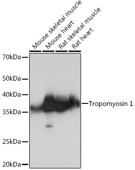
Western blot analysis of various lysates using Tropomyosin 1 Rabbit mAb (CAB8723) at 1:1000 dilution. Secondary antibody: HRP-conjugated Goat anti-Rabbit IgG (H+L) (CABS014) at 1:10000 dilution. Lysates/proteins: 25μg per lane. Blocking buffer: 3% nonfat dry milk in TBST. Detection: ECL Basic Kit (AbGn00020). Exposure time: 3s.
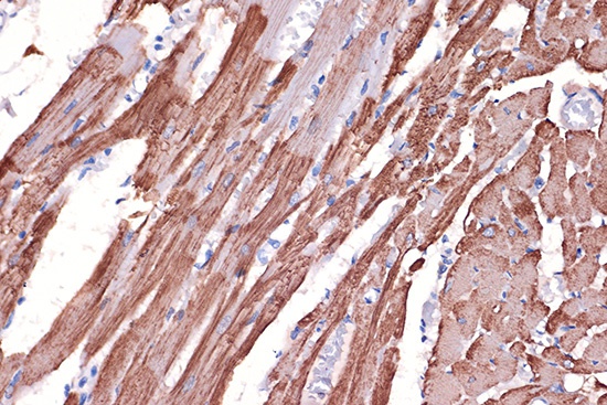
Immunohistochemistry analysis of paraffin-embedded Rat heart using Tropomyosin 1 Rabbit mAb (CAB8723) at dilution of 1:100 (40x lens). Microwave antigen retrieval performed with 0.01M Tris/EDTA Buffer (pH 9.0) prior to IHC staining.
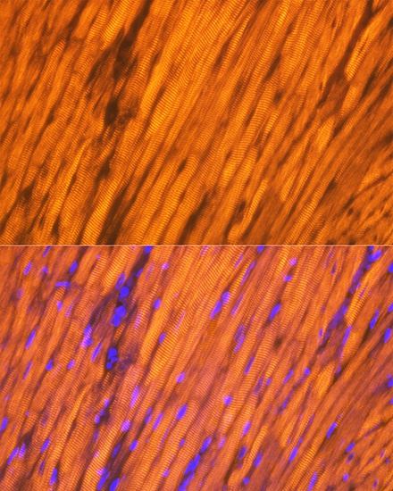
Immunofluorescence analysis of paraffin-embedded rat heart using Tropomyosin 1 Rabbit mAb (CAB8723) at dilution of 1:100 (40x lens). Secondary antibody: Cy3-conjugated Goat anti-Rabbit IgG (H+L) (CABS007) at 1:500 dilution. Blue: DAPI for nuclear staining.
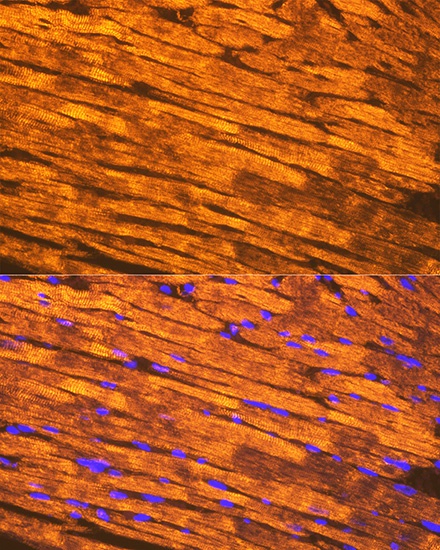
Immunofluorescence analysis of paraffin-embedded mouse heart using Tropomyosin 1 Rabbit mAb (CAB8723) at dilution of 1:100 (40x lens). Secondary antibody: Cy3-conjugated Goat anti-Rabbit IgG (H+L) (CABS007) at 1:500 dilution. Blue: DAPI for nuclear staining.


![Anti-Tropomyosin alpha 1 Chain [R02-5E4] Monoclonal Antibody (AGMB02664)](https://cdn11.bigcommerce.com/s-h68l9z2lnx/images/stencil/590x590/products/273953/676766/anti-tropomyosin-alpha-1-chain-r02-5e4-monoclonal-antibody-agmb02664__59753.1773030500.jpg?c=2 "Anti-Tropomyosin alpha 1 Chain [R02-5E4] Monoclonal Antibody (AGMB02664)")
![Anti-Tropomyosin alpha 1 Chain [R01-3B-6] Monoclonal Antibody (AGMB03911)](https://cdn11.bigcommerce.com/s-h68l9z2lnx/images/stencil/590x590/products/275200/677414/anti-tropomyosin-alpha-1-chain-r01-3b-6-monoclonal-antibody-agmb03911__61979.1773032548.jpg?c=2 "Anti-Tropomyosin alpha 1 Chain [R01-3B-6] Monoclonal Antibody (AGMB03911)")


