Description
Anti-TriMethyl-Histone H3-K27 Antibody (CAB2363)
The Trimethyl-Histone H3 (K27) Polyclonal Antibody (CAB2363) is a key tool for researchers studying epigenetic modifications in gene expression. This antibody, raised in rabbits, specifically targets the trimethylated form of histone H3 at lysine 27, a crucial histone modification involved in gene regulation and chromatin remodeling. Histone modifications, such as trimethylation of histone H3 at lysine 27, play a critical role in controlling gene expression and cellular functions. The CAB2363 antibody is highly reactive with human samples and has been validated for use in various applications, including Western blot and immunohistochemistry.
It enables researchers to detect and analyze the presence of trimethylated histone H3 (K27) in different cell types, providing valuable insights into the epigenetic regulation of gene expression.Research into histone modifications like trimethylation of histone H3 at lysine 27 is essential for understanding how changes in chromatin structure can impact gene expression and contribute to various diseases, including cancer and developmental disorders. By studying the role of this specific histone modification, researchers can uncover potential therapeutic targets for intervention in epigenetic-related diseases.
| Antibody Name: | Anti-TriMethyl-Histone H3-K27 Antibody |
| Antibody SKU: | CAB2363 |
| Antibody Size: | 20uL, 50uL, 100uL |
| Application: | WB IHC IF IP ChIP ChIPseq |
| Reactivity: | Human, Mouse, Rat, Other (Wide Range) |
| Host Species: | Rabbit |
| Immunogen: | A synthetic methylated peptide corresponding to residues surrounding K27 of human histone H3 |
| Application: | WB IHC IF IP ChIP ChIPseq |
| Recommended Dilution: | WB 1:500 - 1:2000 IHC 1:50 - 1:200 IF 1:50 - 1:200 IP 1:50 - 1:200 ChIP 1:20 - 1:100 ChIPseq 1:20 - 1:100 |
| Reactivity: | Human, Mouse, Rat, Other (Wide Range) |
| Positive Samples: | HeLa, NIH/3T3 |
| Immunogen: | A synthetic methylated peptide corresponding to residues surrounding K27 of human histone H3 |
| Purification Method: | Affinity purification |
| Storage Buffer: | Store at -20'C. Avoid freeze / thaw cycles. Buffer: PBS with 0.02% sodium azide, 50% glycerol, pH7.3. |
| Isotype: | IgG |
| Sequence: | Email for sequence |
| Gene ID: | 8290 |
| Uniprot: | Q16695 |
| Cellular Location: | Chromosome, Nucleus |
| Calculated MW: | 15kDa |
| Observed MW: | 17kDa |
| Synonyms: | H3.4, H3/g, H3FT, H3t, HIST3H3, Histone H3, HIST1H3A |
| Background: | Actin is a key regulator of RNA polymerase (Pol) II-dependent transcription. Positive transcription elongation factor b (P-TEFb), a Cdk9/cyclin T1 heterodimer, has been reported to play a critical role in transcription elongation. However, the relationship between actin and P-TEFb is still not clear. In this study, actin was found to interact with Cdk9, a catalytic subunit of P-TEFb, in elongation complexes. Using immunofluorescence and immunoprecipitation assays, Cdk9 was found to bind to G-actin through the conserved Thr-186 in the T-loop. Overexpression and in vitro kinase assays showed that G-actin promotes P-TEFb-dependent phosphorylation of the Pol II C-terminal domain. An in vitro transcription experiment revealed that the interaction between G-actin and Cdk9 stimulated Pol II transcription elongation. ChIP and immobilized template assays indicated that actin recruited Cdk9 to a transcriptional template in vivo and in vitro. Using cytokine IL-6-inducible p21 gene expression system, we revealed that actin recruited Cdk9 to endogenous gene. Moreover, overexpression of actin and Cdk9 increased histone H3 acetylation and acetylized histone H3 binding to a transcriptional template through the interaction with histone acetyltransferase, p300. Taken together, our results suggested that actin participates in transcription elongation by recruiting Cdk9 for phosphorylation of the Pol II C-terminal domain, and the actin-Cdk9 interaction promotes chromatin remodeling. |
| UniProt Protein Function: | HIST3H3: Core component of nucleosome. Nucleosomes wrap and compact DNA into chromatin, limiting DNA accessibility to the cellular machineries which require DNA as a template. Histones thereby play a central role in transcription regulation, DNA repair, DNA replication and chromosomal stability. DNA accessibility is regulated via a complex set of post-translational modifications of histones, also called histone code, and nucleosome remodeling. The nucleosome is a histone octamer containing two molecules each of H2A, H2B, H3 and H4 assembled in one H3-H4 heterotetramer and two H2A-H2B heterodimers. The octamer wraps approximately 147 bp of DNA. Expressed in testicular cells. Belongs to the histone H3 family. |
| UniProt Protein Details: | Protein type:DNA-binding Chromosomal Location of Human Ortholog: 1q42 Cellular Component: nucleoplasm; nucleus Molecular Function:protein binding; DNA binding; histone binding; protein heterodimerization activity Biological Process: nucleosome assembly; negative regulation of protein oligomerization; protein heterotetramerization; telomere maintenance |
| NCBI Summary: | Histones are basic nuclear proteins that are responsible for the nucleosome structure of the chromosomal fiber in eukaryotes. Nucleosomes consist of approximately 146 bp of DNA wrapped around a histone octamer composed of pairs of each of the four core histones (H2A, H2B, H3, and H4). The chromatin fiber is further compacted through the interaction of a linker histone, H1, with the DNA between the nucleosomes to form higher order chromatin structures. This gene is intronless and encodes a member of the histone H3 family. Transcripts from this gene lack polyA tails; instead, they contain a palindromic termination element. This gene is located separately from the other H3 genes that are in the histone gene cluster on chromosome 6p22-p21.3. [provided by RefSeq, Jul 2008] |
| UniProt Code: | Q16695 |
| NCBI GenInfo Identifier: | 18202512 |
| NCBI Gene ID: | 8290 |
| NCBI Accession: | Q16695.3 |
| UniProt Secondary Accession: | Q16695,Q6FGU4, B2R5K3, |
| UniProt Related Accession: | Q16695 |
| Molecular Weight: | |
| NCBI Full Name: | Histone H3.1t |
| NCBI Synonym Full Names: | histone cluster 3, H3 |
| NCBI Official Symbol: | HIST3H3Â Â |
| NCBI Official Synonym Symbols: | H3t; H3.4; H3/g; H3FTÂ Â |
| NCBI Protein Information: | histone H3.1t; H3/t; histone 3, H3; H3 histone family, member T |
| UniProt Protein Name: | Histone H3.1t |
| UniProt Synonym Protein Names: | H3/g |
| UniProt Gene Name: | HIST3H3Â Â |
| UniProt Entry Name: | H31T_HUMAN |
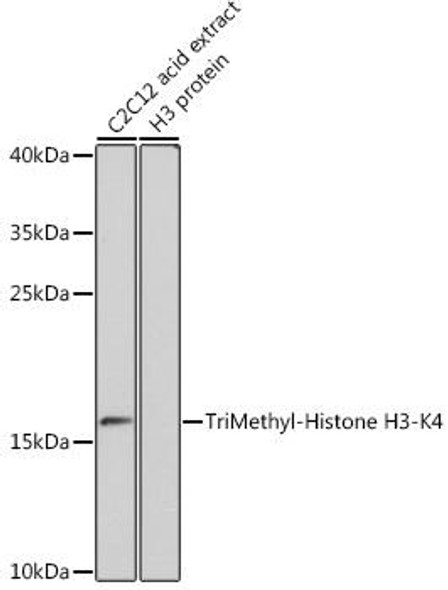
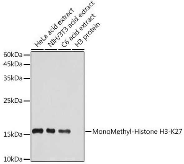
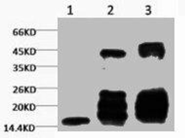
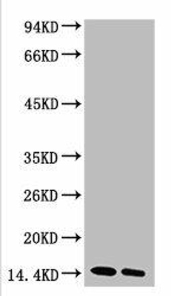
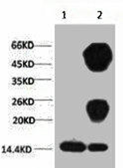
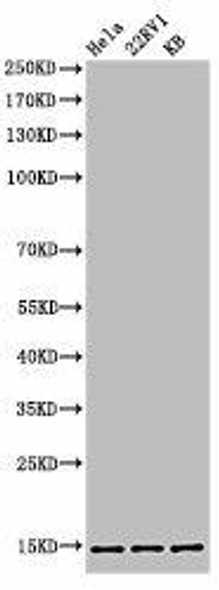
 | Western blot analysis of extracts of various cell lines, using TriMethyl-Histone H3-K27 antibody at 1:1000 dilution. Secondary antibody: HRP Goat Anti-Rabbit IgG (H+L) at 1:10000 dilution. Lysates/proteins: 25ug per lane. Blocking buffer: 3% nonfat dry milk in TBST. Detection: ECL Basic Kit. Exposure time: 60s. |
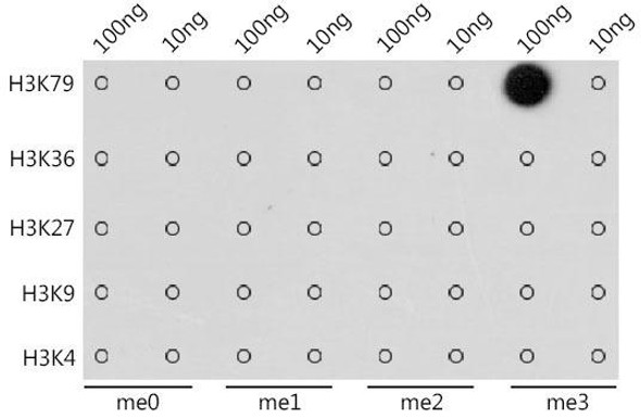
 | Dot-blot analysis of all sorts of methylation peptides using TriMethyl-Histone H3-K27 antibody (A2363). |
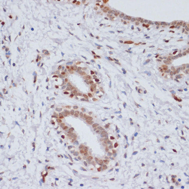
 | Immunohistochemistry of paraffin-embedded rat ovary using TriMethyl-Histone H3-K27 antibody at dilution of 1:100 (40x lens). Perform microwave antigen retrieval with 10 mM PBS buffer pH 7. 2 before commencing with IHC staining protocol. |
 | Immunohistochemistry of paraffin-embedded human breast cancer using TriMethyl-Histone H3-K27 antibody at dilution of 1:100 (40x lens). Perform microwave antigen retrieval with 10 mM PBS buffer pH 7. 2 before commencing with IHC staining protocol. |
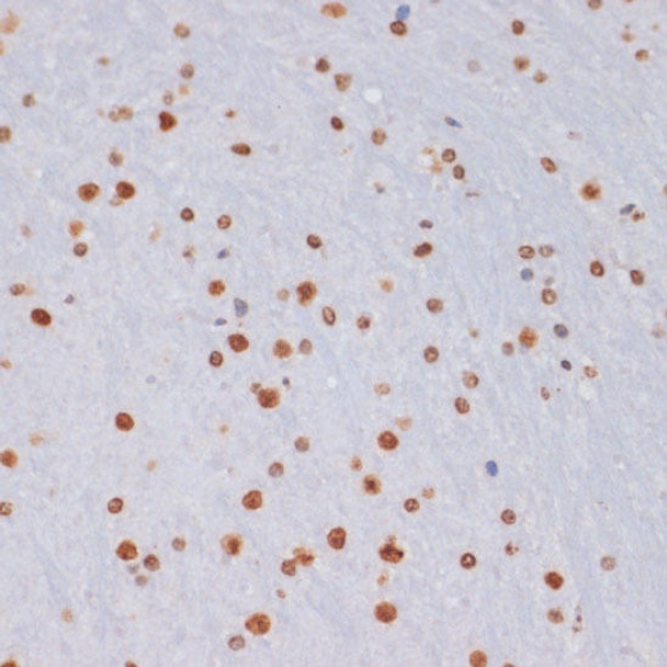
 | Immunohistochemistry of paraffin-embedded mouse brain using TriMethyl-Histone H3-K27 antibody at dilution of 1:100 (40x lens). Perform microwave antigen retrieval with 10 mM PBS buffer pH 7. 2 before commencing with IHC staining protocol. |
 | Immunofluorescence analysis of C6 cells using TriMethyl-Histone H3-K27 antibody at dilution of 1:100. Blue: DAPI for nuclear staining. |
 | Immunofluorescence analysis of NIH/3T3 cells using TriMethyl-Histone H3-K27 antibody at dilution of 1:100. Blue: DAPI for nuclear staining. |
 | Immunofluorescence analysis of U-2 OS cells using TriMethyl-Histone H3-K27 antibody at dilution of 1:100. Blue: DAPI for nuclear staining. |
 | Chromatin immunoprecipitation analysis of extracts of HeLa cells, using TriMethyl-Histone H3-K27 Rabbit pAb antibody and rabbit IgG. The amount of immunoprecipitated DNA was checked by quantitative PCR. Histogram was constructed by the ratios of the immunoprecipitated DNA to the input. |


