The TOM20 Antibody (CAB6774) is a high-quality antibody developed for reliable detection and analysis of target proteins. This antibody, produced in rabbits, exhibits high reactivity with human samples and is validated for use in Western blot applications. By targeting the TOMM20 protein, researchers can accurately detect and analyze its expression in various cell types, making it an essential resource for studies focusing on mitochondrial function and diseases such as neurodegenerative disorders and metabolic syndromes.
This antibody is validated for use in WB, IHC-P, IF/ICC, IP, ELISA applications and has demonstrated reactivity against Human, Mouse, Rat samples.
Product Name:
TOM20 Antibody
SKU:
CAB6774
Size:
20μL, 100μL
Reactivity:
Human, Mouse, Rat
Conjugate:
Unconjugated
Immunogen:
Recombinant protein (or fragment).This information is considered to be commercially sensitive.
Enables protein-transporting ATPase activity and unfolded protein binding activity. Involved in protein targeting to mitochondrion. Located in mitochondria-associated endoplasmic reticulum membrane and mitochondrial outer membrane.
Purification Method
Affinity purification
Gene ID
9804
RRID
AB_2767357
Buffer Information
Store at -20℃. Avoid freeze / thaw cycles. Buffer: PBS containing 50% glycerol, preserved with proclin300 or sodium azide, pH 7.3.
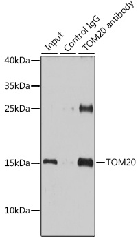
Immunoprecipitation analysis of 200 μg extracts of HeLa cells using 3 μg TOM20 antibody (CAB6774). Western blot was performed from the immunoprecipitate using TOM20 antibody (CAB6774) at a dilution of 1:1000.
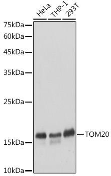
Western blot analysis of various lysates using [KO Validated] TOM20 Rabbit pAb (CAB6774) at 1:3000 dilution. Secondary antibody: HRP-conjugated Goat anti-Rabbit IgG (H+L) (CABS014) at 1:10000 dilution. Lysates/proteins: 25μg per lane. Blocking buffer: 3% nonfat dry milk in TBST. Detection: ECL Basic Kit (AbGn00020). Exposure time: 1s.
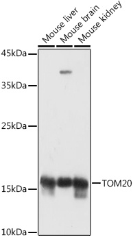
Western blot analysis of various lysates using [KD Validated] TOM20 Rabbit pAb (CAB6774) at 1:1000 dilution. Secondary antibody: HRP-conjugated Goat anti-Rabbit IgG (H+L) (CABS014) at 1:10000 dilution. Lysates/proteins: 25μg per lane. Blocking buffer: 3% nonfat dry milk in TBST. Detection: ECL Basic Kit (AbGn00020). Exposure time: 1s.
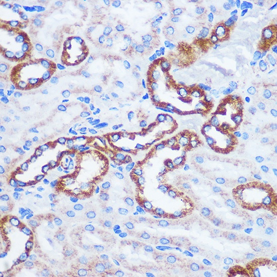
Immunohistochemistry analysis of paraffin-embedded Mouse kidney using TOM20 Rabbit pAb (CAB6774) at dilution of 1:100 (40x lens). Microwave antigen retrieval performed with 0.01M PBS Buffer (pH 7.2) prior to IHC staining.
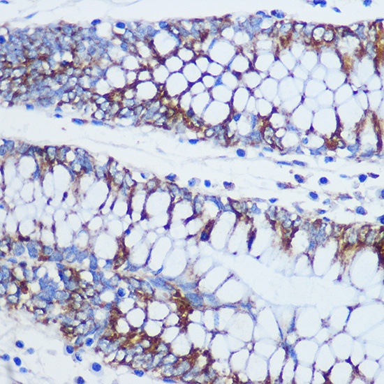
Immunohistochemistry analysis of paraffin-embedded Human colon using TOM20 Rabbit pAb (CAB6774) at dilution of 1:100 (40x lens). Microwave antigen retrieval performed with 0.01M PBS Buffer (pH 7.2) prior to IHC staining.
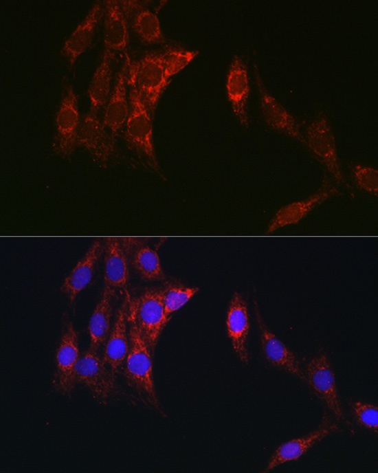
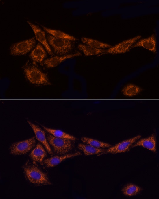
Immunofluorescence analysis of C6 cells using [KO Validated] TOM20 Rabbit pAb (CAB6774) at dilution of 1:100 (40x lens). Secondary antibody: Cy3-conjugated Goat anti-Rabbit IgG (H+L) (CABS007) at 1:500 dilution. Blue: DAPI for nuclear staining.
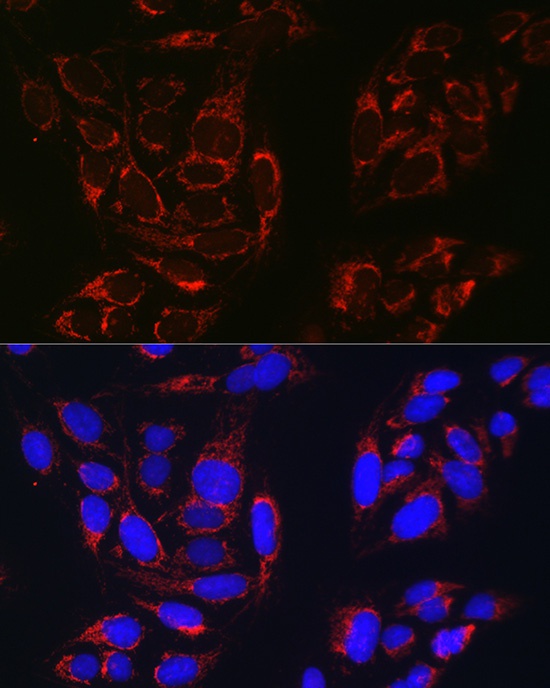
Immunofluorescence analysis of U-2 OS cells using [KO Validated] TOM20 Rabbit pAb (CAB6774) at dilution of 1:100 (40x lens). Secondary antibody: Cy3-conjugated Goat anti-Rabbit IgG (H+L) (CABS007) at 1:500 dilution. Blue: DAPI for nuclear staining.
Immunofluorescence analysis of C6 cells using [KO Validated] TOM20 Rabbit pAb (CAB6774) at dilution of 1:100 (40x lens). Secondary antibody: Cy3-conjugated Goat anti-Rabbit IgG (H+L) (CABS007) at 1:500 dilution. Blue: DAPI for nuclear staining.
Immunofluorescence analysis of U2OS cells using [KO Validated] TOM20 Rabbit pAb (CAB6774) at dilution of 1:100 (40x lens). Secondary antibody: Cy3-conjugated Goat anti-Rabbit IgG (H+L) (CABS007) at 1:500 dilution. Blue: DAPI for nuclear staining.
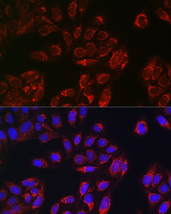
Immunofluorescence analysis of HeLa cells using TOM20 Rabbit pAb (CAB6774) at dilution of 1:200 (40x lens). Secondary antibody: Cy3-conjugated Goat anti-Rabbit IgG (H+L) (CABS007) at 1:500 dilution. Blue: DAPI for nuclear staining.
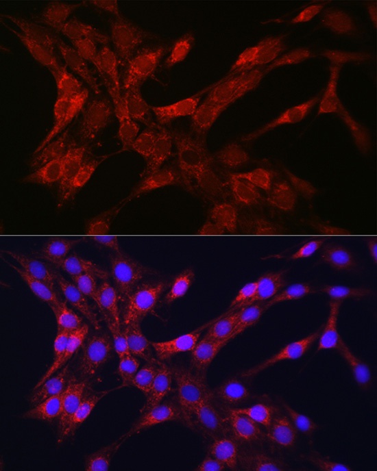
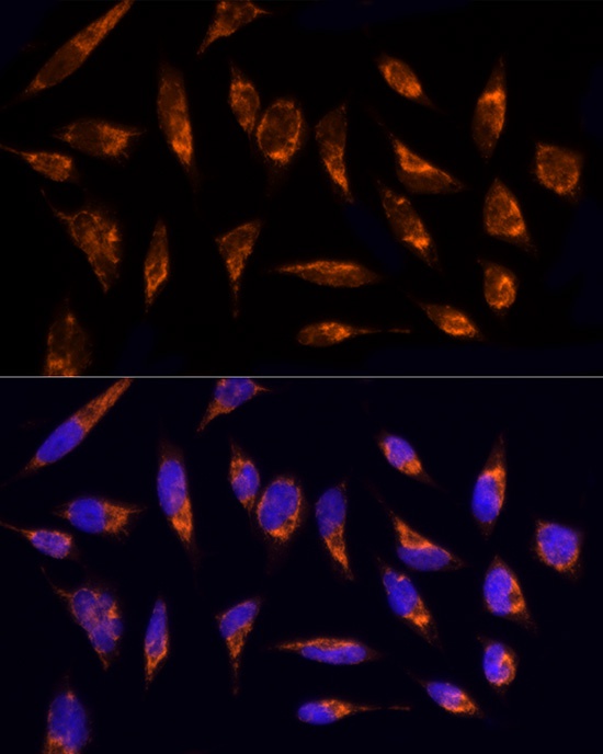
Immunofluorescence analysis of PC-12 cells using TOM20 Rabbit pAb (CAB6774) at dilution of 1:200 (40x lens). Secondary antibody: Cy3-conjugated Goat anti-Rabbit IgG (H+L) (CABS007) at 1:500 dilution. Blue: DAPI for nuclear staining.