The SPK Antibody (CAB4852) is a high-quality antibody developed for reliable detection and analysis of target proteins. This antibody, produced in rabbits, exhibits high specificity and sensitivity towards human samples, making it an excellent choice for experiments such as Western blot analysis. By targeting the SPK protein, this antibody allows for precise detection and characterization in a variety of cell types, making it ideal for investigations in immunology and cancer research.
This antibody is validated for use in WB, IHC-P, ELISA applications and has demonstrated reactivity against Human, Rat samples.
Product Name:
SPK Antibody
SKU:
CAB4852
Size:
20μL, 100μL
Reactivity:
Human, Rat
Conjugate:
Unconjugated
Immunogen:
Recombinant protein (or fragment).This information is considered to be commercially sensitive.
Recommended starting concentration is 1 μg/mL. Please optimize the concentration based on your specific assay requirements.
Synonyms:
SPK, CT84, TOPK, HEL164, Nori-3
Positive Sample:
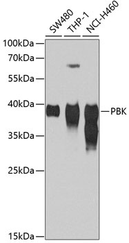
SW480, THP-1, NCI-H460
Cellular Localization:
Nucleus.
Calculated MW:
36kDa
Observed MW:
36kDa
This gene encodes a serine/threonine protein kinase related to the dual specific mitogen-activated protein kinase kinase (MAPKK) family. Evidence suggests that mitotic phosphorylation is required for its catalytic activity. The encoded protein may be involved in the activation of lymphoid cells and support testicular functions, with a suggested role in the process of spermatogenesis. Overexpression of this gene has been implicated in tumorigenesis. Alternative splicing results in multiple transcript variants.
Purification Method
Affinity purification
Gene ID
55872
RRID
AB_2765905
Buffer Information
Store at -20℃. Avoid freeze / thaw cycles. Buffer: PBS containing 50% glycerol, preserved with proclin300 or sodium azide, pH 7.3.
Western blot analysis of various lysates using SPK Rabbit pAb (CAB4852) at 1:1000 dilution. Secondary antibody: HRP-conjugated Goat anti-Rabbit IgG (H+L) (CABS014) at 1:10000 dilution. Lysates/proteins: 25μg per lane. Blocking buffer: 3% nonfat dry milk in TBST. Detection: ECL Basic Kit (AbGn00020). Exposure time: 90s.
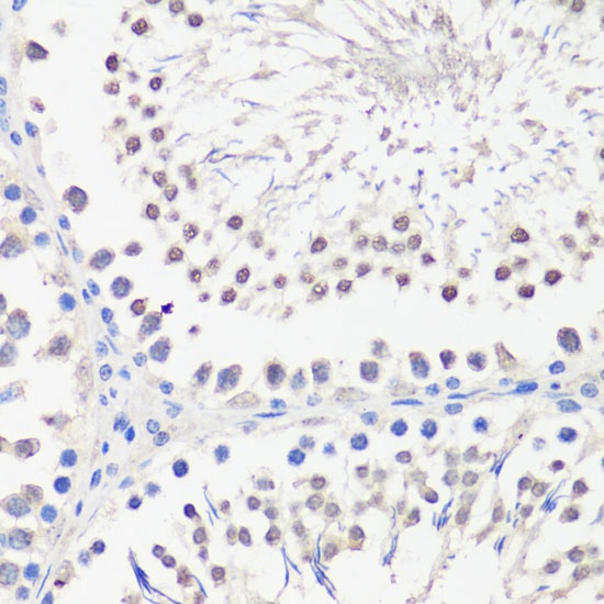
Immunohistochemistry analysis of paraffin-embedded Rat testis using SPK Rabbit pAb (CAB4852) at dilution of 1:100 (40x lens). Microwave antigen retrieval performed with 0.01M PBS Buffer (pH 7.2) prior to IHC staining.