The SOS2 Antibody (PAC063399) is a high-quality research tool designed for studying the SOS2 protein, a key regulator in the Ras signaling pathway. This polyclonal antibody, generated in rabbits, exhibits strong reactivity with human samples and is validated for use in Western blot applications.SOS2 is involved in cell growth, differentiation, and survival, making it a crucial component in cell signaling processes. Dysregulation of the Ras pathway, in which SOS2 plays a pivotal role, is commonly associated with cancer development and progression.
By targeting SOS2 with this antibody, researchers can gain insights into its function and potential as a therapeutic target in cancer research.The ability of the SOS2 Antibody to specifically bind to the SOS2 protein enables precise detection and analysis in various cell types, supporting investigations in oncology and cell biology. Understanding the role of SOS2 in signaling pathways is essential for advancing knowledge of cancer biology and developing targeted treatments for a variety of malignancies.
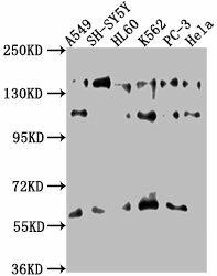
Western Blot. Positive WB detected in: A549 whole cell lysate, SH-SY5Y whole cell lysate, HL60 whole cell lysate, K562 whole cell lysate, PC-3 whole cell lysate, Hela whole cell lysate. All lanes: SOS2 antibody at 1:1000. Secondary. Goat polyclonal to rabbit IgG at 1/50000 dilution. Predicted band size: 153, 150 kDa. Observed band size: 153 kDa.
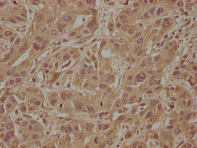
IHC image of PACO63399 diluted at 1:100 and staining in paraffin-embedded human liver cancer performed on a Leica BondTM system. After dewaxing and hydration, antigen retrieval was mediated by high pressure in a citrate buffer (pH 6.0). Section was blocked with 10% normal goat serum 30min at RT. Then primary antibody (1% BSA) was incubated at 4°C overnight. The primary is detected by a biotinylated secondary antibody and visualized using an HRP conjugated SP system.
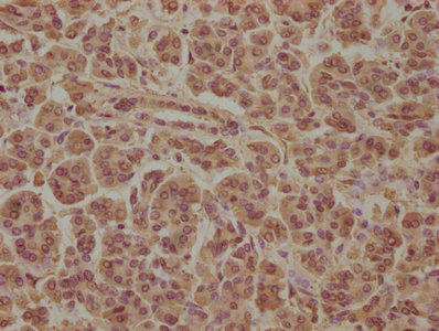
IHC image of PACO63399 diluted at 1:100 and staining in paraffin-embedded human pancreatic tissue performed on a Leica BondTM system. After dewaxing and hydration, antigen retrieval was mediated by high pressure in a citrate buffer (pH 6.0). Section was blocked with 10% normal goat serum 30min at RT. Then primary antibody (1% BSA) was incubated at 4°C overnight. The primary is detected by a biotinylated secondary antibody and visualized using an HRP conjugated SP system.
Background:
Promotes the exchange of Ras-bound GDP by GTP.
Synonyms:
Son of sevenless homolog 2 (SOS-2), SOS2
UniProt Protein Function:
SOS2: a guanine nucleotide exchange factor that activates Ras by catalyzing the exchange of bound GDP for GTP. Interacts with GRB2.Protein type: Motility/polarity/chemotaxis; GEFs, Ras; GEFs; Adaptor/scaffoldChromosomal Location of Human Ortholog: 14q21Cellular Component: cytosolMolecular Function: Rho guanyl-nucleotide exchange factor activity; protein binding; DNA binding; protein heterodimerization activityBiological Process: positive regulation of small GTPase mediated signal transduction; axon guidance; regulation of small GTPase mediated signal transduction; nerve growth factor receptor signaling pathway; positive regulation of apoptosis; small GTPase mediated signal transduction


.")
.")
. Section was blocked with 10% normal goat serum 30min at RT. Then primary antibody (1% BSA) was incubated at 4°C overnight. The primary is detected by a Goat anti-rabbit IgG labeled by HRP and visualized using 0.05% DAB.")
.")

.")