The SMAD5 Monoclonal Antibody is a valuable tool for researchers studying the SMAD5 protein, a key player in the TGF-β signaling pathway. This antibody, produced in mice, exhibits high specificity and sensitivity for detecting SMAD5 in human samples, making it ideal for use in Western blot and immunohistochemistry applications.SMAD5 is a transcription factor that plays a crucial role in cell growth, differentiation, and development. Dysregulation of the TGF-β signaling pathway, in which SMAD5 is involved, has been linked to various diseases, including cancer, fibrosis, and developmental disorders. By targeting SMAD5 with this monoclonal antibody, researchers can gain insights into the mechanisms underlying these conditions and potentially identify new therapeutic targets.
With its ability to specifically bind to SMAD5, this monoclonal antibody enables the accurate detection and quantification of the protein in different cell types and tissues. Its reliability and consistency make it a valuable tool for advancing research in the fields of molecular biology, cell biology, and cancer research. Gain a deeper understanding of SMAD5 function and its role in disease pathology with the SMAD5 Monoclonal Antibody.
Product Name:
Smad5 Monoclonal Antibody
SKU:
CAB22749
Size:
20uL, 100uL
Isotype:
IgG
Host Species:
Rabbit
Reactivity:
Human,Rat
Immunogen:
Recombinant fusion protein containing a sequence corresponding to amino acids 186-260 of human Smad5 (NP_005894.3).
The protein encoded by this gene is involved in the transforming growth factor beta signaling pathway that results in an inhibition of the proliferation of hematopoietic progenitor cells. The encoded protein is activated by bone morphogenetic proteins type 1 receptor kinase, and may be involved in cancer. Alternative splicing results in multiple transcript variants.
Purification Method:
Affinity purification
Gene ID:
4090
Clone Number:
ARC55344
Storage Buffer:
Store at -20℃. Avoid freeze / thaw cycles.Buffer: PBS with 0.05% proclin300,0.05% BSA,50% glycerol,pH7.3.
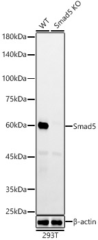
Western blot analysis of lysates from wild type(WT) and Smad5 knockout (KO) 293T(KO) cells, using [KO Validated] Smad5 Rabbit mAb (CAB22749) at 1:1000 dilution.Secondary antibody: HRP Goat Anti-Rabbit IgG (H+L) (CABS014) at 1:10000 dilution.Lysates/proteins: 25μg per lane.Blocking buffer: 3% nonfat dry milk in TBST.Detection: ECL Enhanced Kit (AbGn00021).Exposure time: 180s.
 and Smad5 knockout (KO) 293T(KO) cells, using Smad5 antibody (CAB22749) at 1:1000 dilution. Secondary antibody: HRP Goat Anti-Rabbit IgG (H+L) at 1:10000 dilution. Lysates/proteins: 25μg per lane. Blocking buffer: 3% nonfat dry milk in TBST.")
 and Smad5 knockout (KO) 293T(KO) cells, using Smad5 antibody (CAB22749) at 1:1000 dilution. Secondary antibody: HRP Goat Anti-Rabbit IgG (H+L) at 1:10000 dilution. Lysates/proteins: 25μg per lane. Blocking buffer: 3% nonfat dry milk in TBST.")


")