The SIRPB1 Polyclonal Antibody (PAC059133) is a valuable tool for researchers studying SIRPB1, a cell surface molecule known for its role in immune regulation. This antibody, generated in rabbits, shows high specificity and reactivity with human samples, making it an ideal choice for Western blot applications. By binding to the SIRPB1 protein, this antibody allows for accurate detection and analysis in a variety of cell types, making it a crucial tool for studies in immunology and cancer research.
SIRPB1, also known as signal-regulatory protein beta 1, is involved in modulating immune responses and has been implicated in various diseases, including cancer and autoimmune disorders. By understanding the function of SIRPB1, researchers can gain insights into potential therapeutic targets for these conditions. The SIRPB1 Polyclonal Antibody is a key component in advancing our understanding of immune regulation and its potential impact on disease progression and treatment strategies.
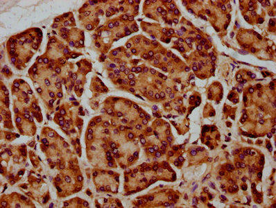
IHC image of PACO59133 diluted at 1:400 and staining in paraffin-embedded human pancreatic cancer performed on a Leica BondTM system. After dewaxing and hydration, antigen retrieval was mediated by high pressure in a citrate buffer (pH 6.0). Section was blocked with 10% normal goat serum 30min at RT. Then primary antibody (1% BSA) was incubated at 4°C overnight. The primary is detected by a biotinylated secondary antibody and visualized using an HRP conjugated SP system.
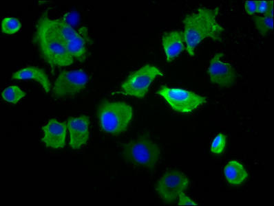
Immunofluorescence staining of MCF-7 cells with PACO59133 at 1:133, counter-stained with DAPI. The cells were fixed in 4% formaldehyde, permeabilized using 0.2% Triton X-100 and blocked in 10% normal Goat Serum. The cells were then incubated with the antibody overnight at 4°C. The secondary antibody was Alexa Fluor 488-congugated AffiniPure Goat Anti-Rabbit IgG(H+L).
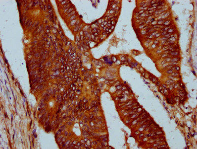
IHC image of PACO59133 diluted at 1:400 and staining in paraffin-embedded human colon cancer performed on a Leica BondTM system. After dewaxing and hydration, antigen retrieval was mediated by high pressure in a citrate buffer (pH 6.0). Section was blocked with 10% normal goat serum 30min at RT. Then primary antibody (1% BSA) was incubated at 4°C overnight. The primary is detected by a biotinylated secondary antibody and visualized using an HRP conjugated SP system.
Background:
Immunoglobulin-like cell surface receptor involved in the negative regulation of receptor tyrosine kinase-coupled signaling processes.
Synonyms:
Signal-regulatory protein beta-1 isoform 3 (SIRP-beta-1 isoform 3), SIRPB1
UniProt Protein Function:
SIRPB1 iso3: Immunoglobulin-like cell surface receptor involved in the negative regulation of receptor tyrosine kinase-coupled signaling processes. Participates also in the recruitment of tyrosine kinase SYK. 3 isoforms of the human protein are produced by alternative splicing.Protein type: Membrane protein, integralChromosomal Location of Human Ortholog: 20p13Cellular Component: integral to plasma membrane; plasma membraneMolecular Function: protein bindingBiological Process: cell surface receptor linked signal transduction; innate immune response; signal transduction
UniProt Protein Details:
NCBI Summary:
The protein encoded by this gene is a member of the signal-regulatory-protein (SIRP) family, and also belongs to the immunoglobulin superfamily. SIRP family members are receptor-type transmembrane glycoproteins known to be involved in the negative regulation of receptor tyrosine kinase-coupled signaling processes. This protein was found to interact with TYROBP/DAP12, a protein bearing immunoreceptor tyrosine-based activation motifs. This protein was also reported to participate in the recruitment of tyrosine kinase SYK. Multiple transcript variants encoding different isoforms have been found for this gene. [provided by RefSeq, Feb 2009]


")

 (RPES1605)")