The SARDH Antibody (CAB9541) is a high-quality antibody developed for reliable detection and analysis of target proteins. This antibody, generated in rabbits, exhibits high reactivity with human samples and has been validated for use in Western blot applications.SARDH plays a crucial role in the detoxification of sulfite, a byproduct of sulfur metabolism that can be toxic if not properly metabolized. Dysregulation of SARDH activity has been implicated in various metabolic disorders and diseases, making it a target of interest in research related to metabolic diseases, oxidative stress, and neurological disorders.
This antibody is validated for use in WB, IF/ICC, ELISA applications and has demonstrated reactivity against Mouse, Rat samples.
Product Name:
SARDH Antibody
SKU:
CAB9541
Size:
20μL, 100μL
Reactivity:
Mouse, Rat
Conjugate:
Unconjugated
Immunogen:
Recombinant protein (or fragment).This information is considered to be commercially sensitive.
Recommended starting concentration is 1 μg/mL. Please optimize the concentration based on your specific assay requirements.
Synonyms:
SAR, SDH, SARD, BPR-2, DMGDHL1, SARDH
Positive Sample:
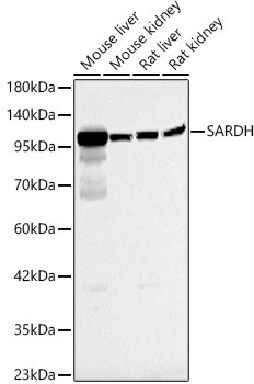
Mouse liver, Mouse kidney, Rat liver, Rat kidney
Cellular Localization:
Mitochondrion Matrix.
Calculated MW:
101kDa
Observed MW:
100kDa
This gene encodes an enzyme localized to the mitochondrial matrix which catalyzes the oxidative demethylation of sarcosine. This enzyme is distinct from another mitochondrial matrix enzyme, dimethylglycine dehydrogenase, which catalyzes a reaction resulting in the formation of sarcosine. Mutations in this gene are associated with sarcosinemia. Alternatively spliced transcript variants have been described.
Purification Method
Affinity purification
Gene ID
1757
RRID
AB_2772143
Buffer Information
Store at -20℃. Avoid freeze / thaw cycles. Buffer: PBS containing 50% glycerol, preserved with proclin300 or sodium azide, pH 7.3.
Western blot analysis of various lysates, using SARDH Rabbit pAb (CAB9541) at 1:1000 dilution. Secondary antibody: HRP-conjugated Goat anti-Rabbit IgG (H+L) (CABS014) at 1:10000 dilution. Lysates/proteins: 25μg per lane. Blocking buffer: 3% nonfat dry milk in TBST. Detection: ECL Basic Kit (AbGn00020). Exposure time: 40s.
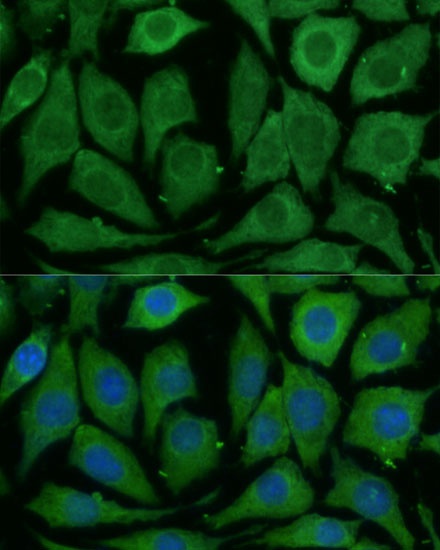
Immunofluorescence analysis of L929 cells using SARDH Rabbit pAb (CAB9541) at dilution of 1:100 (40x lens). Secondary antibody: Cy3-conjugated Goat anti-Rabbit IgG (H+L) (CABS007) at 1:500 dilution. Blue: DAPI for nuclear staining.