The RTN4RL2 Antibody (PAC062059) is a high-quality polyclonal antibody designed for research involving RTN4RL2, a protein involved in various cellular processes, including cell migration, proliferation, and differentiation. This antibody, produced in rabbits, is highly specific and reactive with human samples, making it a valuable tool for studying the function of RTN4RL2 in different cellular contexts.Validated for use in Western blot applications, this antibody binds specifically to the RTN4RL2 protein, allowing for accurate detection and analysis in a range of cell types.
Its high specificity and sensitivity make it ideal for immunology and cancer research studies, where RTN4RL2 has been implicated in tumor progression, metastasis, and drug resistance.With its involvement in regulating key cellular processes, RTN4RL2 is a promising target for investigations into cancer biology and therapeutic development. By understanding the function of RTN4RL2, researchers can potentially identify new treatment strategies for cancer and other diseases where RTN4RL2 plays a role.
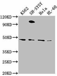
Western Blot. Positive WB detected in: K562 whole cell lysate, SH-SY5Y whole cell lysate, Hela whole cell lysate, HL-60 whole cell lysate. All lanes: RTN4RL2 antibody at 1:2000. Secondary. Goat polyclonal to rabbit IgG at 1/50000 dilution. Predicted band size: 47, 22 kDa. Observed band size: 47 kDa.
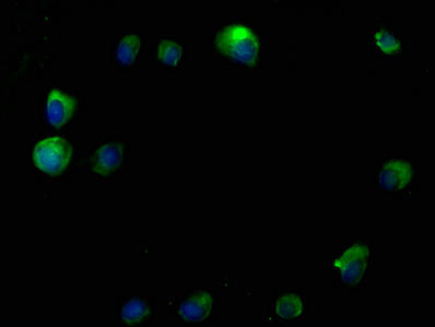
Immunofluorescence staining of MCF-7 cells with PACO62059 at 1:50, counter-stained with DAPI. The cells were fixed in 4% formaldehyde, permeabilized using 0.2% Triton X-100 and blocked in 10% normal Goat Serum. The cells were then incubated with the antibody overnight at 4°C. The secondary antibody was Alexa Fluor 488-congugated AffiniPure Goat Anti-Rabbit IgG(H+L).
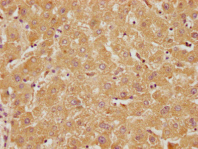
IHC image of PACO62059 diluted at 1:100 and staining in paraffin-embedded human liver tissue performed on a Leica BondTM system. After dewaxing and hydration, antigen retrieval was mediated by high pressure in a citrate buffer (pH 6.0). Section was blocked with 10% normal goat serum 30min at RT. Then primary antibody (1% BSA) was incubated at 4°C overnight. The primary is detected by a biotinylated secondary antibody and visualized using an HRP conjugated SP system.
Background:
Cell surface receptor that plays a functionally redundant role in the inhibition of neurite outgrowth mediated by MAG (By similarity). Plays a functionally redundant role in postnatal brain development. Contributes to normal axon migration across the brain midline and normal formation of the corpus callosum. Does not seem to play a significant role in regulating axon regeneration in the adult central nervous system. Protects motoneurons against apoptosis; protection against apoptosis is probably mediated by MAG (By similarity). Like other family members, plays a role in restricting the number dendritic spines and the number of synapses that are formed during brain development. Signaling mediates activation of Rho and downstream reorganization of the actin cytoskeleton.
RTN4RL2: May play a role in regulating axonal regeneration and plasticity in the adult central nervous system. Belongs to the Nogo receptor family.Protein type: Membrane protein, GPI anchor; Cell development/differentiation; Receptor, misc.Chromosomal Location of Human Ortholog: 11q12.1Cellular Component: anchored to plasma membrane; cell surface; cytoplasmMolecular Function: protein kinase inhibitor activity; receptor activityBiological Process: axon regeneration; cytokine and chemokine mediated signaling pathway; negative regulation of JAK-STAT cascade; negative regulation of protein kinase activity


. Section was blocked with 10% normal goat serum 30min at RT. Then primary antibody (1% BSA) was incubated at 4°C overnight. The primary is detected by a Goat anti-rabbit IgG labeled by HRP and visualized using 0.05% DAB.")
.")

.")

. Section was blocked with 10% normal goat serum 30min at RT. Then primary antibody (1% BSA) was incubated at 4°C overnight. The primary is detected by a Goat anti-rabbit polymer IgG labeled by HRP and visualized using 0.05% DAB.")
. Section was blocked with 10% normal goat serum 30min at RT. Then primary antibody (1% BSA) was incubated at 4°C overnight. The primary is detected by a Goat anti-rabbit polymer IgG labeled by HRP and visualized using 0.05% DAB.")

.")