Rat bFGF / FGF2 / BFGF / FGF2 ELISA Kit
- SKU:
- RTFI00595
- Product Type:
- ELISA Kit
- Size:
- 96 Assays
- Uniprot:
- P13109
- Sensitivity:
- 9.375pg/ml
- Range:
- 15.625-1000pg/ml
- ELISA Type:
- Sandwich
- Synonyms:
- bFGF, FGF2, FGF-2, FGF2AS, GFG1, HBGH-2, NUDT6, Prostatropin, Basic fibroblast growth factor, basic fibroblast growth factor bFGF, FGFB, fibroblast growth factor 2, basic, HBGF-2, heparin-binding growth factor 2
- Reactivity:
- Rat
- Research Area:
- Cardiovascular

Description
Rat PDGFB ELISA Kit
Basic fibroblast growth factor (bFGF), also known as FGF2 (Fibroblast Growth Factor 2), is a protein that plays a crucial role in cell growth, development, and tissue repair.The Rat bFGF/FGF2 ELISA Kit is specifically designed for the quantitative measurement of rat basic fibroblast growth factor (bFGF/FGF2) in biological samples. The kit aids in various research applications, including studying the role of bFGF/FGF2 in development, tissue repair, angiogenesis, and disease processes.
Key Features
| Save Time | Pre-coated 96 well plate | |
| Quick Start | Kit includes all necessary reagents | |
| Publication Ready | Reproducible and reliable results |
Overview
|
Product Name: |
Rat bFGF/FGF2 (Basic Fibroblast Growth Factor) ELISA Kit |
|
Product Code: |
RTFI00595 |
|
Size: |
96 Assays |
|
Alias: |
bFGF, FGF2, FGF-2, FGF2AS, GFG1, HBGH-2, NUDT6, Prostatropin, Basic fibroblast growth factor, basic fibroblast growth factor bFGF, FGFB, fibroblast growth factor 2, basic, HBGF-2, heparin-binding growth factor 2 |
|
Detection Method: |
Sandwich ELISA, Double Antibody |
|
Reactivity: |
Rat |
|
Sensitivity: |
9.375pg/ml |
|
Range: |
15.625-1000pg/ml |
|
Storage: |
4°C for 6 months |
|
Note: |
For Research Use Only |
Additional Information
|
Recovery |
Matrices listed below were spiked with certain level of Rat bFGF/FGF2 and the recovery rates were calculated by comparing the measured value to the expected amount of Rat bFGF/FGF2 in samples.
|
||||||||||||||||||||
|
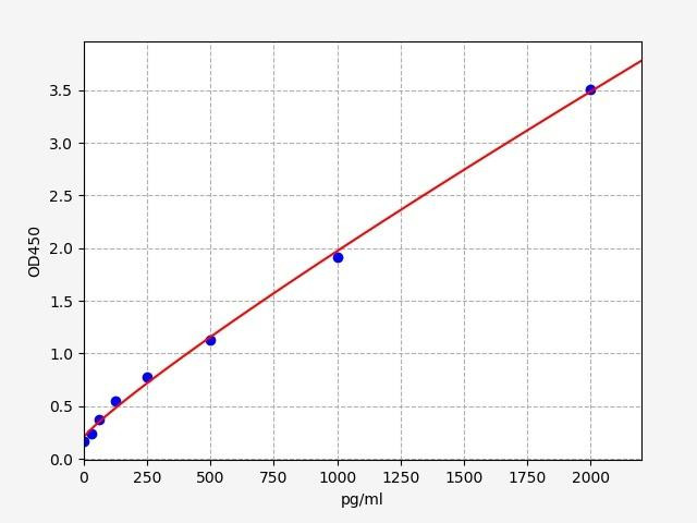
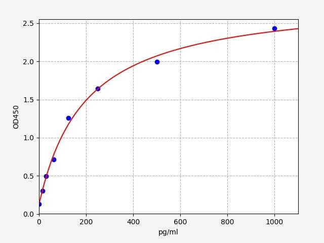
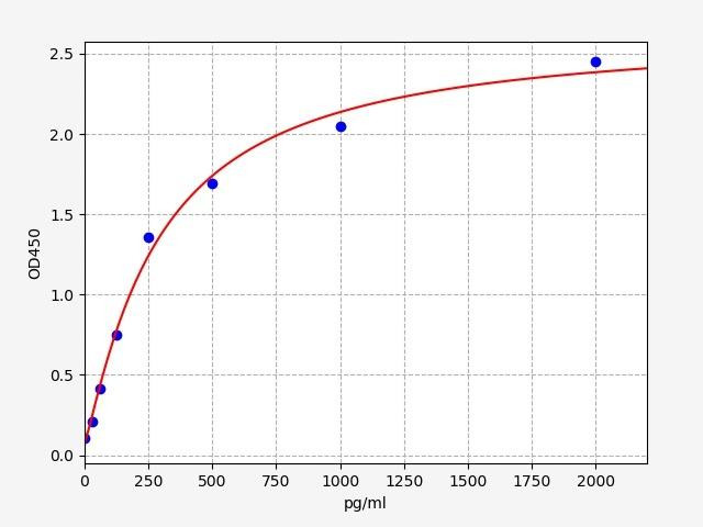
Linearity: |
The linearity of the kit was assayed by testing samples spiked with appropriate concentration of Rat bFGF/FGF2 and their serial dilutions. The results were demonstrated by the percentage of calculated concentration to the expected.
|
||||||||||||||||||||
|
CV(%) |
Intra Assay <8 Inter Assay <10 |
Kit Components
| Component | Quantity | Storage |
|
ELISA Microplate (Dismountable) |
8x12 strips |
2-8°C/-20°C |
|
Lyophilized Standard |
2 |
2-8°C/-20°C |
|
Sample/Standard Dlution Buffer |
20ml |
2-8°C |
|
Biotin-labeled Antibody (Concentrated) |
120ul |
2-8°C (Protection from light) |
|
Antibody Dilution Buffer |
10ml |
2-8°C |
|
HRP-Streptavidin Conjugate (SABC) |
120ul |
2-8°C(Protect from light) |
|
SABC Dilution Buffer |
10ml |
2-8°C |
|
TMB Substrate |
10ml |
2-8°C(Protection from light) |
|
Stop Solution |
10ml |
2-8°C |
|
Wash Buffer (25X) |
30ml |
2-8°C |
|
Plate Sealer |
5 |
- |
Other materials required:
- Microplate reader with 450 nm wavelength filter
- Multichannel Pipette, Pipette, microcentrifuge tubes and disposable pipette tips
- Incubator
- Deionized or distilled water
- Absorbent paper
- Buffer resevoir
Protocol
*Note: Protocols are specific to each batch/lot. For the exact instructions please follow the protocol included in your kit.
Before adding to wells, equilibrate the SABC working solution and TMB substrate for at least 30 min at 37°C. When diluting samples and reagents, they must be mixed completely and evenly. It is recommended to plot a standard curve for each test.
| Step | Procedure |
|
1. |
Set standard, test sample and control (zero) wells on the pre-coated plate respectively, and then, record their positions. It is recommended to measure each standard and sample in duplicate. Wash plate 2 times before adding standard, sample and control (zero) wells! |
|
2. |
Aliquot 0.1ml standard solutions into the standard wells. |
|
3. |
Add 0.1 ml of Sample / Standard dilution buffer into the control (zero) well. |
|
4. |
Add 0.1 ml of properly diluted sample ( Human serum, plasma, tissue homogenates and other biological fluids.) into test sample wells. |
|
5. |
Seal the plate with a cover and incubate at 37 °C for 90 min. |
|
6. |
Remove the cover and discard the plate content, clap the plate on the absorbent filter papers or other absorbent material. Do NOT let the wells completely dry at any time. Wash plate X2. |
|
7. |
Add 0.1 ml of Biotin- detection antibody working solution into the above wells (standard, test sample & zero wells). Add the solution at the bottom of each well without touching the side wall. |
|
8. |
Seal the plate with a cover and incubate at 37°C for 60 min. |
|
9. |
Remove the cover, and wash plate 3 times with Wash buffer. Let wash buffer rest in wells for 1 min between each wash. |
|
10. |
Add 0.1 ml of SABC working solution into each well, cover the plate and incubate at 37°C for 30 min. |
|
11. |
Remove the cover and wash plate 5 times with Wash buffer, and each time let the wash buffer stay in the wells for 1-2 min. |
|
12. |
Add 90 µl of TMB substrate into each well, cover the plate and incubate at 37°C in dark within 10-20 min. (Note: This incubation time is for reference use only, the optimal time should be determined by end user.) And the shades of blue can be seen in the first 3-4 wells (with most concentrated standard solutions), the other wells show no obvious color. |
|
13. |
Add 50 µl of Stop solution into each well and mix thoroughly. The color changes into yellow immediately. |
|
14. |
Read the O.D. absorbance at 450 nm in a microplate reader immediately after adding the stop solution. |
Sample Type
When carrying out an ELISA assay it is important to prepare your samples in order to achieve the best possible results. Below we have a list of procedures for the preparation of samples for different sample types.
| Sample Type | Protocol |
|
Serum |
If using serum separator tubes, allow samples to clot for 30 minutes at room temperature. Centrifuge for 10 minutes at 1,000x g. Collect the serum fraction and assay promptly or aliquot and store the samples at -80°C. Avoid multiple freeze-thaw cycles. If serum separator tubes are not being used, allow samples to clot overnight at 2-8°C. Centrifuge for 10 minutes at 1,000x g. Remove serum and assay promptly or aliquot and store the samples at -80°C. Avoid multiple freeze-thaw cycles. |
|
Plasma |
Collect plasma using EDTA or heparin as an anticoagulant. Centrifuge samples at 4°C for 15 mins at 1000 × g within 30 mins of collection. Collect the plasma fraction and assay promptly or aliquot and store the samples at -80°C. Avoid multiple freeze-thaw cycles. Note: Over haemolysed samples are not suitable for use with this kit. |
|
Urine & Cerebrospinal Fluid |
Collect the urine (mid-stream) in a sterile container, centrifuge for 20 mins at 2000-3000 rpm. Remove supernatant and assay immediately. If any precipitation is detected, repeat the centrifugation step. A similar protocol can be used for cerebrospinal fluid. |
|
Cell culture supernatant |
Collect the cell culture media by pipette, followed by centrifugation at 4°C for 20 mins at 1500 rpm. Collect the clear supernatant and assay immediately. |
|
Cell lysates |
Solubilize cells in lysis buffer and allow to sit on ice for 30 minutes. Centrifuge tubes at 14,000 x g for 5 minutes to remove insoluble material. Aliquot the supernatant into a new tube and discard the remaining whole cell extract. Quantify total protein concentration using a total protein assay. Assay immediately or aliquot and store at ≤ -20 °C. |
|
Tissue homogenates |
The preparation of tissue homogenates will vary depending upon tissue type. Rinse tissue with 1X PBS to remove excess blood & homogenize in 20ml of 1X PBS (including protease inhibitors) and store overnight at ≤ -20°C. Two freeze-thaw cycles are required to break the cell membranes. To further disrupt the cell membranes you can sonicate the samples. Centrifuge homogenates for 5 mins at 5000xg. Remove the supernatant and assay immediately or aliquot and store at -20°C or -80°C. |
|
Tissue lysates |
Rinse tissue with PBS, cut into 1-2 mm pieces, and homogenize with a tissue homogenizer in PBS. Add an equal volume of RIPA buffer containing protease inhibitors and lyse tissues at room temperature for 30 minutes with gentle agitation. Centrifuge to remove debris. Quantify total protein concentration using a total protein assay. Assay immediately or aliquot and store at ≤ -20 °C |
|
Breast Milk |
Collect milk samples and centrifuge at 10,000 x g for 60 min at 4°C. Aliquot the supernatant and assay. For long term use, store samples at -80°C. Minimize freeze/thaw cycles. |
bFGF/ FGF2 Background
basic Fibroblast Growth Factor/ Fibroblast growth Factor 2
Basic fibroblast growth factor (bFGF), also known as FGF2 (Fibroblast Growth Factor 2), is a protein that plays a crucial role in cell growth, development, and tissue repair. It belongs to the fibroblast growth factor family, which comprises a group of signaling molecules involved in various physiological processes.
bFGF/FGF2 Gene
The rat Fgf2 gene is located on chromosome 2 (2q25). It consists of multiple exons and introns, similar to the human gene. Alternative splicing of the Fgf2 gene in rats, as in humans, generates different isoforms of bFGF. These isoforms have variations in their biological activities and tissue distribution.
bFGF/FGF2 Structure
Structurally, bFGF is a small protein consisting of 155 amino acids. It contains a characteristic core domain, formed by a beta-trefoil structure, and two short alpha-helical segments. The protein lacks a signal peptide, which means it is not secreted through the conventional endoplasmic reticulum-Golgi pathway. Instead, it can be released from cells through non-classical pathways or by binding to extracellular matrix components.
Predicted Structure of Rat FGF2. Source: Uniprot
bFGF/FGF2 Function
The primary function of bFGF is to regulate cell proliferation, differentiation, migration, and survival. It acts as a paracrine and autocrine factor, influencing a wide range of cell types, including fibroblasts, endothelial cells, smooth muscle cells, and various progenitor cells. bFGF exerts its effects by binding to specific cell surface receptors, known as fibroblast growth factor receptors (FGFRs), which activate downstream signaling pathways, such as the MAPK/ERK and PI3K/Akt pathways.
bFGF is involved in numerous physiological and pathological processes. During embryonic development, it participates in the formation of various organs and tissues, including the central nervous system, limb buds, and blood vessels. In adults, bFGF plays a critical role in tissue repair and wound healing by stimulating cell proliferation and angiogenesis. Additionally, it is involved in tissue regeneration and has been investigated for its potential therapeutic applications in conditions such as myocardial infarction, stroke, and peripheral arterial disease.
FGF2 Signalling Pathways
The signaling pathway of basic fibroblast growth factor 2 (FGF2) involves the binding of FGF2 to its specific cell surface receptors, known as fibroblast growth factor receptors (FGFRs). Upon binding, the FGF2-FGFR complex undergoes conformational changes, leading to the activation of intracellular signaling cascades. The main pathways involved in FGF2 signaling include the mitogen-activated protein kinase/extracellular signal-regulated kinase (MAPK/ERK) pathway ( cell proliferation, differentiation and survival)and the phosphoinositide 3-kinase/protein kinase B (PI3K/Akt) pathway (proliferation, migration, angiogenesis and tissue development).
Besides these major pathways, FGF2 signaling can also activate other signaling cascades, including the signal transducer and activator of transcription (STAT) pathway and the protein kinase C (PKC) pathway. These pathways contribute to the diverse effects of FGF2 on cell behavior, including proliferation, migration, angiogenesis, and tissue development.
FGF2 and Cancer
Clinically, bFGF has garnered significant attention due to its potential implications in cancer. It can promote tumor growth by stimulating angiogenesis, allowing tumors to receive nutrients and oxygen for sustained growth. Moreover, bFGF has been associated with metastasis and is considered a prognostic marker in several types of cancer. As a result, targeting bFGF and its signaling pathways has become an area of interest in anticancer research, with the aim of developing therapies that can inhibit its effects on tumor growth and progression.
Rat bFGF/FGF2 ELISA Kit FAQs
Q: What is the purpose of the Rat bFGF/FGF2 ELISA Kit?
The Rat bFGF/FGF2 ELISA Kit is specifically designed for the quantitative measurement of rat basic fibroblast growth factor (bFGF/FGF2) in biological samples. The kit aids in various research applications, including studying the role of bFGF/FGF2 in development, tissue repair, angiogenesis, and disease processes.
Q: What types of samples can be used with this kit?
The Rat bFGF/FGF2 ELISA Kit is suitable for a wide range of sample types, including serum, plasma, cell lysates, tissue homogenates, and other rat-derived samples. It is important to follow the provided sample preparation instructions to ensure accurate and reliable results.
Q: Can the Rat bFGF/FGF2 ELISA kit be used for diagnostic purposes?
No, the Rat bFGF/FGF2 ELISA Kit is intended for research purposes only and is not approved for diagnostic or clinical use. It provides quantitative data for research applications and should not be used for medical diagnosis or patient management.
Q: Where can I find additional technical support or assistance with the Rat bFGF/FGF2 ELISA kit?
For any technical inquiries or assistance regarding the Rat bFGF/FGF2 ELISA kit, you can reach out to our team. They will be available to answer your questions and provide the necessary guidance to ensure a successful experiment.
Related Products
| Rat VEGF ELISA Kit | |
|---|---|
| ELISA TYPE: | Sandwich ELISA, Double Antibody |
| SENSITIVITY: | 18.75pg/ml |
| RANGE: | 31.25-2000pg/ml |
| Rat FGF1 / FGF acidic ELISA Kit | |
|---|---|
| SENSITIVITY: | 9.375pg/ml |
| RANGE: | 15.625-1000pg/ml |
| ELISA TYPE: | Sandwich ELISA, Double Antibody |
| Rat PDGFB ELISA Kit | |
|---|---|
| ELISA TYPE: | Sandwich ELISA, Double Antibody |
| SENSITIVITY: | 18.75pg/ml |
| RANGE: | 31.25-2000pg/ml |
Related Products

Rabbit bFGF / FGF2 / Basic Fibroblast Growth Factor ELISA Kit

Porcine bFGF / FGF2 / Basic Fibroblast Growth Factor ELISA Kit

Chicken bFGF / FGF2 / Basic Fibroblast Growth Factor ELISA Kit

