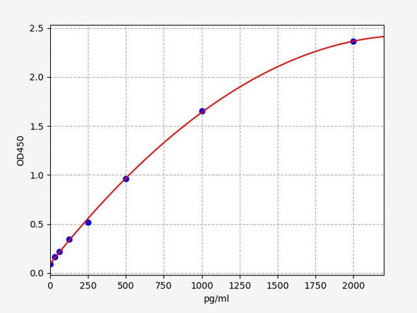
Rat 14-3-3 protein zeta/delta (Ywhaz) ELISA Kit (RTEB0957)
- SKU:
- RTEB0957
- Product Type:
- ELISA Kit
- Size:
- 96 Assays
- Uniprot:
- P63102
- Range:
- 31.2-2000 pg/mL
- ELISA Type:
- Sandwich
- Synonyms:
- Ywhaz, Msfs1, Mitochondrial import stimulation factor S1 subunit, Protein kinase C inhibitor protein 1, KCIP-1, 14-3-3 protein zeta, delta
- Reactivity:
- Rat
Description
Rat 14-3-3 protein zeta/delta (Ywhaz) ELISA Kit
The Rat 14-3-3 Protein Zeta/Delta (YWHAZ) ELISA Kit is specifically designed for the accurate quantification of 14-3-3 protein zeta/delta levels in rat serum, plasma, and cell culture supernatants. This kit offers high sensitivity and specificity, ensuring precise and reliable results for a variety of research applications.14-3-3 protein zeta/delta is a key regulatory protein involved in various cellular processes, including cell signaling, apoptosis, and cell cycle control.
Dysregulation of this protein has been linked to numerous diseases, making it a valuable biomarker for understanding disease mechanisms and potential therapeutic targets.With easy-to-follow protocols and minimal sample volume requirements, the Rat 14-3-3 Protein Zeta/Delta (YWHAZ) ELISA Kit is a versatile tool for researchers studying the role of 14-3-3 protein zeta/delta in health and disease. Order yours today to advance your research efforts.
| Product Name: | Rat 14-3-3 protein zeta/delta (Ywhaz) ELISA Kit |
| SKU: | RTEB0957 |
| Size: | 96T |
| Target: | Rat 14-3-3 protein zeta/delta (Ywhaz) |
| Synonyms: | Mitochondrial import stimulation factor S1 subunit, Protein kinase C inhibitor protein 1, KCIP-1, Msfs1 |
| Assay Type: | Sandwich |
| Detection Method: | ELISA |
| Reactivity: | Rat |
| Detection Range: | 31.2-2000pg/mL |
| Sensitivity: | 15.68pg/mL |
| Intra CV: | 4.1% | ||||||||||||||||||||
| Inter CV: | 7.4% | ||||||||||||||||||||
| Linearity: |
| ||||||||||||||||||||
| Recovery: |
| ||||||||||||||||||||
| Function: | Adapter protein implicated in the regulation of a large spectrum of both general and specialized signaling pathways. Binds to a large number of partners, usually by recognition of a phosphoserine or phosphothreonine motif. Binding generally results in the modulation of the activity of the binding partner. |
| Uniprot: | P63102 |
| Sample Type: | Serum, plasma, tissue homogenates, cell culture supernates and other biological fluids |
| Specificity: | Natural and recombinant rat 14-3-3 protein zeta/delta |
| Sub Unit: | Homodimer. Heterodimerizes with YWHAE (By similarity). Homo- and hetero-dimerization is inhibited by phosphorylation on Ser-58 (By similarity). Interacts with FOXO4, NOXA1, SSH1 and ARHGEF2. Interacts with CDK16 and with WEE1 (C-terminal). Interacts with MLF1 (phosphorylated form); the interaction retains it in the cytoplasm. Interacts with Thr-phosphorylated ITGB2. Interacts with Pseudomonas aeruginosa exoS (unphosphorylated form). Interacts with BAX; the interaction occurs in the cytoplasm. Under stress conditions, MAPK8-mediated phosphorylation releases BAX to mitochondria. Interacts with phosphorylated RAF1; the interaction is inhibited when YWHAZ is phosphorylated on Thr-232. Interacts with TP53; the interaction enhances p53 transcriptional activity. The Ser-58 phosphorylated form inhibits this interaction and p53 transcriptional activity. Interacts with ABL1 (phosphorylated form); the interaction retains ABL1 in the cytoplasm. Interacts with PKA-phosphorylated AANAT; the interaction modulates AANAT enzymatic activity by increasing affinity for arylalkylamines and acetyl-CoA and protecting the enzyme from dephosphorylation and proteasomal degradation. It may also prevent thiol-dependent inactivation. Interacts with AKT1; the interaction phosphorylates YWHAZ and modulates dimerization (By similarity). Interacts with GAB2 and SAMSN1. Binds to TLK2 (By similarity). Interacts with BSPRY. Interacts with BCL2L11. Interacts with the 'Thr-369' phosphorylated form of DAPK2 (By similarity). Interacts with PI4KB, TBC1D22A and TBC1D22B (By similarity). Interacts with ZFP36L1 (via phosphorylated form); this interaction occurs in a p38 MAPK- and AKT-signaling pathways (By similarity). Interacts with SLITRK1. |
| Research Area: | Neurosciences |
| Subcellular Location: | Cytoplasm Melanosome Located to stage I to stage IV melanosomes. |
| Storage: | Please see kit components below for exact storage details |
| Note: | For research use only |
| UniProt Protein Function: | 14-3-3 zeta: a protein of the 14-3-3 family of proteins which mediate signal transduction by binding to phosphoserine-containing proteins. A multifunctional regulator of the cell signaling processes. Phosphorylation apparently disrupts homodimerization. |
| UniProt Protein Details: | Protein type:Adaptor/scaffold; Motility/polarity/chemotaxis Chromosomal Location of Human Ortholog: 7q22 Cellular Component: extracellular space; focal adhesion; leading edge; mitochondrion; nucleus; perinuclear region of cytoplasm; postsynaptic density; protein complex; vesicle Molecular Function:cadherin binding; identical protein binding; protein binding; protein complex binding; protein domain specific binding; protein kinase binding; RNA binding; transcription factor binding; ubiquitin protein ligase binding Biological Process: establishment of Golgi localization; histamine secretion by mast cell; protein targeting; protein targeting to mitochondrion; response to drug |
| NCBI Summary: | 30kDa component of the mitochondrial import stimulation factor, a protein complex that facilitates the import of in vitro synthesized precursor proteins into mitochondria [RGD, Feb 2006] |
| UniProt Code: | P63102 |
| NCBI GenInfo Identifier: | 62990183 |
| NCBI Gene ID: | 25578 |
| NCBI Accession: | NP_037143.2 |
| UniProt Secondary Accession: | P63102,P35215, P70197, P97286, Q52KK1, Q6IRF4, |
| UniProt Related Accession: | P63102 |
| Molecular Weight: | 27,771 Da |
| NCBI Full Name: | 14-3-3 protein zeta/delta |
| NCBI Synonym Full Names: | tyrosine 3-monooxygenase/tryptophan 5-monooxygenase activation protein, zeta |
| NCBI Official Symbol: | Ywhaz |
| NCBI Official Synonym Symbols: | 14-3-3z |
| NCBI Protein Information: | 14-3-3 protein zeta/delta |
| UniProt Protein Name: | 14-3-3 protein zeta/delta |
| UniProt Synonym Protein Names: | Mitochondrial import stimulation factor S1 subunit; Protein kinase C inhibitor protein 1; KCIP-1 |
| Protein Family: | 14-3-3 protein |
| UniProt Gene Name: | Ywhaz |
| Component | Quantity (96 Assays) | Storage |
| ELISA Microplate (Dismountable) | 8×12 strips | -20°C |
| Lyophilized Standard | 2 | -20°C |
| Sample Diluent | 20ml | -20°C |
| Assay Diluent A | 10mL | -20°C |
| Assay Diluent B | 10mL | -20°C |
| Detection Reagent A | 120µL | -20°C |
| Detection Reagent B | 120µL | -20°C |
| Wash Buffer | 30mL | 4°C |
| Substrate | 10mL | 4°C |
| Stop Solution | 10mL | 4°C |
| Plate Sealer | 5 | - |
Other materials and equipment required:
- Microplate reader with 450 nm wavelength filter
- Multichannel Pipette, Pipette, microcentrifuge tubes and disposable pipette tips
- Incubator
- Deionized or distilled water
- Absorbent paper
- Buffer resevoir
*Note: The below protocol is a sample protocol. Protocols are specific to each batch/lot. For the correct instructions please follow the protocol included in your kit.
Allow all reagents to reach room temperature (Please do not dissolve the reagents at 37°C directly). All the reagents should be mixed thoroughly by gently swirling before pipetting. Avoid foaming. Keep appropriate numbers of strips for 1 experiment and remove extra strips from microtiter plate. Removed strips should be resealed and stored at -20°C until the kits expiry date. Prepare all reagents, working standards and samples as directed in the previous sections. Please predict the concentration before assaying. If values for these are not within the range of the standard curve, users must determine the optimal sample dilutions for their experiments. We recommend running all samples in duplicate.
| Step | |
| 1. | Add Sample: Add 100µL of Standard, Blank, or Sample per well. The blank well is added with Sample diluent. Solutions are added to the bottom of micro ELISA plate well, avoid inside wall touching and foaming as possible. Mix it gently. Cover the plate with sealer we provided. Incubate for 120 minutes at 37°C. |
| 2. | Remove the liquid from each well, don't wash. Add 100µL of Detection Reagent A working solution to each well. Cover with the Plate sealer. Gently tap the plate to ensure thorough mixing. Incubate for 1 hour at 37°C. Note: if Detection Reagent A appears cloudy warm to room temperature until solution is uniform. |
| 3. | Aspirate each well and wash, repeating the process three times. Wash by filling each well with Wash Buffer (approximately 400µL) (a squirt bottle, multi-channel pipette,manifold dispenser or automated washer are needed). Complete removal of liquid at each step is essential. After the last wash, completely remove remaining Wash Buffer by aspirating or decanting. Invert the plate and pat it against thick clean absorbent paper. |
| 4. | Add 100µL of Detection Reagent B working solution to each well. Cover with the Plate sealer. Incubate for 60 minutes at 37°C. |
| 5. | Repeat the wash process for five times as conducted in step 3. |
| 6. | Add 90µL of Substrate Solution to each well. Cover with a new Plate sealer and incubate for 10-20 minutes at 37°C. Protect the plate from light. The reaction time can be shortened or extended according to the actual color change, but this should not exceed more than 30 minutes. When apparent gradient appears in standard wells, user should terminatethe reaction. |
| 7. | Add 50µL of Stop Solution to each well. If color change does not appear uniform, gently tap the plate to ensure thorough mixing. |
| 8. | Determine the optical density (OD value) of each well at once, using a micro-plate reader set to 450 nm. User should open the micro-plate reader in advance, preheat the instrument, and set the testing parameters. |
| 9. | After experiment, store all reagents according to the specified storage temperature respectively until their expiry. |
When carrying out an ELISA assay it is important to prepare your samples in order to achieve the best possible results. Below we have a list of procedures for the preparation of samples for different sample types.
| Sample Type | Protocol |
| Serum | If using serum separator tubes, allow samples to clot for 30 minutes at room temperature. Centrifuge for 10 minutes at 1,000x g. Collect the serum fraction and assay promptly or aliquot and store the samples at -80°C. Avoid multiple freeze-thaw cycles. If serum separator tubes are not being used, allow samples to clot overnight at 2-8°C. Centrifuge for 10 minutes at 1,000x g. Remove serum and assay promptly or aliquot and store the samples at -80°C. Avoid multiple freeze-thaw cycles. |
| Plasma | Collect plasma using EDTA or heparin as an anticoagulant. Centrifuge samples at 4°C for 15 mins at 1000 × g within 30 mins of collection. Collect the plasma fraction and assay promptly or aliquot and store the samples at -80°C. Avoid multiple freeze-thaw cycles. Note: Over haemolysed samples are not suitable for use with this kit. |
| Urine & Cerebrospinal Fluid | Collect the urine (mid-stream) in a sterile container, centrifuge for 20 mins at 2000-3000 rpm. Remove supernatant and assay immediately. If any precipitation is detected, repeat the centrifugation step. A similar protocol can be used for cerebrospinal fluid. |
| Cell culture supernatant | Collect the cell culture media by pipette, followed by centrifugation at 4°C for 20 mins at 1500 rpm. Collect the clear supernatant and assay immediately. |
| Cell lysates | Solubilize cells in lysis buffer and allow to sit on ice for 30 minutes. Centrifuge tubes at 14,000 x g for 5 minutes to remove insoluble material. Aliquot the supernatant into a new tube and discard the remaining whole cell extract. Quantify total protein concentration using a total protein assay. Assay immediately or aliquot and store at ≤ -20 °C. |
| Tissue homogenates | The preparation of tissue homogenates will vary depending upon tissue type. Rinse tissue with 1X PBS to remove excess blood & homogenize in 20ml of 1X PBS (including protease inhibitors) and store overnight at ≤ -20°C. Two freeze-thaw cycles are required to break the cell membranes. To further disrupt the cell membranes you can sonicate the samples. Centrifuge homogenates for 5 mins at 5000xg. Remove the supernatant and assay immediately or aliquot and store at -20°C or -80°C. |
| Tissue lysates | Rinse tissue with PBS, cut into 1-2 mm pieces, and homogenize with a tissue homogenizer in PBS. Add an equal volume of RIPA buffer containing protease inhibitors and lyse tissues at room temperature for 30 minutes with gentle agitation. Centrifuge to remove debris. Quantify total protein concentration using a total protein assay. Assay immediately or aliquot and store at ≤ -20 °C. |
| Breast Milk | Collect milk samples and centrifuge at 10,000 x g for 60 min at 4°C. Aliquot the supernatant and assay. For long term use, store samples at -80°C. Minimize freeze/thaw cycles. |


