Rabbit IgG (Immunoglobulin G) ELISA Kit (RBFI00157)
- SKU:
- RBFI00157
- Product Type:
- ELISA Kit
- Size:
- 96 Assays
- Sensitivity:
- 0.938ng/ml
- Range:
- 1.563-100ng/ml
- ELISA Type:
- Sandwich
- Synonyms:
- IgG, Immunoglobulin G
- Reactivity:
- Rabbit

Description
Rabbit IgG (Immunoglobulin G) ELISA Kit
IgG, or immunoglobulin G, is an antibody found in the blood and bodily fluids. It plays a critical role in the immune system's defense against pathogens. These Rabbit IgG ELISA Kits are specifically designed for research purposes to detect and quantify rabbit IgG antibodies and alloe. Researchers to study the immune response in rabbits, assess antibody levels during vaccine development, monitor diseases or infections, and evaluate the effectiveness of experimental treatments or interventions in rabbit models.
Key Features
| Save Time | Pre-coated 96 well plate | |
| Quick Start | Kit includes all necessary reagents | |
| Publication Ready | Reproducible and reliable results |
Overview
|
Product Name: |
Rabbit IgG (Immunoglobulin G) ELISA Kit |
|
Product Code: |
RBFI00157 |
|
Size: |
96 Assays |
|
Alias: |
IgG ELISA Kit, Immunoglobulin G ELISA Kit |
|
Detection Method: |
Sandwich ELISA, Double Antibody |
|
Reactivity: |
Rabbit |
|
Sensitivity: |
< 0.938ng/ml |
|
Range: |
1.563-100ng/ml |
|
Storage: |
4°C for 6 months |
|
Note: |
For Research Use Only |
Additional Information
|
Recovery |
Matrices listed below were spiked with certain level of Rabbit IgG (Immunoglobulin G) and the recovery rates were calculated by comparing the measured value to the expected amount of Rabbit IgG (Immunoglobulin G) in samples. Please contact us for more information. |
|
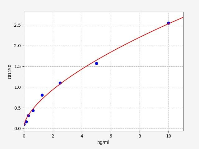
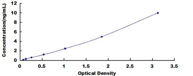
Linearity: |
The linearity of the kit was assayed by testing samples spiked with appropriate concentration of Rabbit IgG (Immunoglobulin G) and their serial dilutions. The results were demonstrated by the percentage of calculated concentration to the expected. Please contact us for more information. |
|
CV(%) |
Intra Assay <8 Inter Assay <10 |
Kit Components
| Component | Quantity | Storage |
|
ELISA Microplate (Dismountable) |
8x12 strips |
2-8°C/-20°C |
|
Lyophilized Standard |
2 |
2-8°C/-20°C |
|
Sample/Standard Dlution Buffer |
20ml |
2-8°C |
|
Biotin-labeled Antibody (Concentrated) |
120ul |
2-8°C (Protection from light) |
|
Antibody Dilution Buffer |
10ml |
2-8°C |
|
HRP-Streptavidin Conjugate (SABC) |
120ul |
2-8°C (Protect from light) |
|
SABC Dilution Buffer |
10ml |
2-8°C |
|
TMB Substrate |
10ml |
2-8°C (Protection from light) |
|
Stop Solution |
10ml |
2-8°C |
|
Wash Buffer (25X) |
30ml |
2-8°C |
|
Plate Sealer |
5 |
- |
Other materials required:
- Microplate reader with 450 nm wavelength filter
- Multichannel Pipette, Pipette, microcentrifuge tubes and disposable pipette tips
- Incubator
- Deionized or distilled water
- Absorbent paper
- Buffer resevoir
Protocol
*Note: Protocols are specific to each batch/lot. For the exact instructions please follow the protocol included in your kit.
Before adding to wells, equilibrate the SABC working solution and TMB substrate for at least 30 min at 37°C. When diluting samples and reagents, they must be mixed completely and evenly. It is recommended to plot a standard curve for each test.
| Step | Procedure |
|
1. |
Set standard, test sample and control (zero) wells on the pre-coated plate respectively, and then, record their positions. It is recommended to measure each standard and sample in duplicate. Wash plate 2 times before adding standard, sample and control (zero) wells! |
|
2. |
Aliquot 0.1ml standard solutions into the standard wells. |
|
3. |
Add 0.1 ml of Sample / Standard dilution buffer into the control (zero) well. |
|
4. |
Add 0.1 ml of properly diluted sample ( Human serum, plasma, tissue homogenates and other biological fluids.) into test sample wells. |
|
5. |
Seal the plate with a cover and incubate at 37 °C for 90 min. |
|
6. |
Remove the cover and discard the plate content, clap the plate on the absorbent filter papers or other absorbent material. Do NOT let the wells completely dry at any time. Wash plate X2. |
|
7. |
Add 0.1 ml of Biotin- detection antibody working solution into the above wells (standard, test sample & zero wells). Add the solution at the bottom of each well without touching the side wall. |
|
8. |
Seal the plate with a cover and incubate at 37°C for 60 min. |
|
9. |
Remove the cover, and wash plate 3 times with Wash buffer. Let wash buffer rest in wells for 1 min between each wash. |
|
10. |
Add 0.1 ml of SABC working solution into each well, cover the plate and incubate at 37°C for 30 min. |
|
11. |
Remove the cover and wash plate 5 times with Wash buffer, and each time let the wash buffer stay in the wells for 1-2 min. |
|
12. |
Add 90 µl of TMB substrate into each well, cover the plate and incubate at 37°C in dark within 10-20 min. (Note: This incubation time is for reference use only, the optimal time should be determined by end user.) And the shades of blue can be seen in the first 3-4 wells (with most concentrated standard solutions), the other wells show no obvious color. |
|
13. |
Add 50 µl of Stop solution into each well and mix thoroughly. The color changes into yellow immediately. |
|
14. |
Read the O.D. absorbance at 450 nm in a microplate reader immediately after adding the stop solution. |
Sample Preparation
When carrying out an ELISA assay it is important to prepare your samples in order to achieve the best possible results. Below we have a list of procedures for the preparation of samples for different sample types.
| Sample Type | Protocol |
|
Serum |
If using serum separator tubes, allow samples to clot for 30 minutes at room temperature. Centrifuge for 10 minutes at 1,000x g. Collect the serum fraction and assay promptly or aliquot and store the samples at -80°C. Avoid multiple freeze-thaw cycles. If serum separator tubes are not being used, allow samples to clot overnight at 2-8°C. Centrifuge for 10 minutes at 1,000x g. Remove serum and assay promptly or aliquot and store the samples at -80°C. Avoid multiple freeze-thaw cycles. |
|
Plasma |
Collect plasma using EDTA or heparin as an anticoagulant. Centrifuge samples at 4°C for 15 mins at 1000 × g within 30 mins of collection. Collect the plasma fraction and assay promptly or aliquot and store the samples at -80°C. Avoid multiple freeze-thaw cycles. Note: Over haemolysed samples are not suitable for use with this kit. |
|
Urine & Cerebrospinal Fluid |
Collect the urine (mid-stream) in a sterile container, centrifuge for 20 mins at 2000-3000 rpm. Remove supernatant and assay immediately. If any precipitation is detected, repeat the centrifugation step. A similar protocol can be used for cerebrospinal fluid. |
|
Cell culture supernatant |
Collect the cell culture media by pipette, followed by centrifugation at 4°C for 20 mins at 1500 rpm. Collect the clear supernatant and assay immediately. |
|
Cell lysates |
Solubilize cells in lysis buffer and allow to sit on ice for 30 minutes. Centrifuge tubes at 14,000 x g for 5 minutes to remove insoluble material. Aliquot the supernatant into a new tube and discard the remaining whole cell extract. Quantify total protein concentration using a total protein assay. Assay immediately or aliquot and store at ≤ -20 °C. |
|
Tissue homogenates |
The preparation of tissue homogenates will vary depending upon tissue type. Rinse tissue with 1X PBS to remove excess blood & homogenize in 20ml of 1X PBS (including protease inhibitors) and store overnight at ≤ -20°C. Two freeze-thaw cycles are required to break the cell membranes. To further disrupt the cell membranes you can sonicate the samples. Centrifuge homogenates for 5 mins at 5000xg. Remove the supernatant and assay immediately or aliquot and store at -20°C or -80°C. |
|
Tissue lysates |
Rinse tissue with PBS, cut into 1-2 mm pieces, and homogenize with a tissue homogenizer in PBS. Add an equal volume of RIPA buffer containing protease inhibitors and lyse tissues at room temperature for 30 minutes with gentle agitation. Centrifuge to remove debris. Quantify total protein concentration using a total protein assay. Assay immediately or aliquot and store at ≤ -20 °C |
|
Breast Milk |
Collect milk samples and centrifuge at 10,000 x g for 60 min at 4°C. Aliquot the supernatant and assay. For long term use, store samples at -80°C. Minimize freeze/thaw cycles. |
IgG Background
What is IgG?
Immunoglobulin G (IgG) is a vital component of the immune system, belonging to a class of antibodies known as gamma globulins. These antibodies are soluble proteins present in the bloodstream and other bodily fluids, playing a crucial role in defending against harmful pathogens.
IgG Gene and IgG Structure
The immunoglobulin heavy and light chains are coded for by three separate gene families. Each family is located on a separate chromosome - one for the heavy chain and one for each of the light chain types. Each of these gene families has several V region genes and one or more C region genes. The V and C regions genes are not however immediately adjacent to each other. This gene undergoes a process called somatic hypermutation, which generates a diverse array of antibodies with different binding affinities and specificities.
The structure of IgG is composed of four protein chains. These chains include two identical heavy chains and two identical light chains, linked together by disulfide bonds. Each chain comprises variable (V) and constant (C) regions. The V regions contribute to the antigen-binding site, enabling IgG to recognize and attach to specific foreign substances called antigens. The molecular weight of the rabbit IgG is 150kDa for their most common isotype.
IgG Antibody Function
The primary function of IgG is to provide protection against bacterial and viral infections. Plasma cells, derived from activated B cells, are responsible for producing IgG antibodies. These antibodies circulate within the bloodstream and lymphatic system, where they neutralize toxins, enhance the process of phagocytosis (engulfing and destroying pathogens), and facilitate the activation of other immune cells.
IgG Clinical Significance
IgG possesses significant clinical significance. It is the most abundant antibody class in the human body and plays a crucial role in long-term immune memory. The presence or absence of IgG can offer valuable diagnostic insights into an individual's immune response. Elevated levels of IgG may indicate chronic infections, autoimmune diseases, or specific types of cancer. Conversely, reduced levels of IgG can be associated with primary immunodeficiencies or immunosuppression.
IgG Tests
gG tests, also known as IgG antibody tests, are commonly employed to assess an individual's immune response to specific antigens. These tests measure the quantity of IgG antibodies present in the blood, helping determine if a person has been exposed to a particular pathogen or allergen. IgG testing is widely used in diagnosing infections, monitoring vaccine effectiveness, and identifying allergies, including food allergies.
IgG Index
The IgG index, also known as the IgG synthesis rate, is a calculation used in medicine to assess the presence of increased immunoglobulin G (IgG) levels within the central nervous system (CNS). It is primarily used as a diagnostic tool in the evaluation of neurological disorders, particularly those suspected to be associated with inflammation or an immune response.The IgG index is calculated by comparing the concentration of IgG in the cerebrospinal fluid (CSF) to the concentration of IgG in the blood.
An elevated IgG index suggests increased production of IgG within the CNS, indicating a possible immune response or inflammation. This finding can be useful in diagnosing conditions such as multiple sclerosis (MS), neurosyphilis, or other infectious or inflammatory disorders affecting the CNS. However, it is important to note that an elevated IgG index alone is not diagnostic of any specific condition and should be interpreted in conjunction with other clinical findings and diagnostic tests.
IgG and Food Allergies
While IgE antibodies are widely recognized as the primary antibodies involved in immediate allergic reactions, some alternative practitioners and testing laboratories claim that elevated levels of IgG antibodies to specific foods can indicate food intolerances or sensitivities. However, scientific evidence supporting the use of IgG Food sensitivity tests for diagnosing food allergies is limited and inconclusive. Major allergy organizations and medical societies generally do not endorse or recommend IgG testing as a standalone diagnostic tool for food allergies. They argue that the presence of IgG antibodies to a specific food does not necessarily indicate an allergic reaction or intolerance, as IgG antibodies can be present even in individuals without symptoms or clinical significance.
Rabbit IgG ELISA Kit FAQs
Q: What is the purpose of Rabbit IgG ELISA Kits?
Rabbit IgG ELISA Kits are specifically designed to measure the presence and quantity of rabbit IgG antibodies in biological samples. These kits enable researchers to accurately detect and quantify rabbit IgG, which is essential for various applications, including immunology research, diagnostics, and biomedical studies.
Q: What types of samples can be used with Rabbit IgG ELISA Kits?
Rabbit IgG ELISA Kits can be used with a variety of biological samples, including serum, plasma, cell culture supernatants, and other bodily fluids. However, it is important to follow the specific instructions provided with the kit, as the compatibility of different sample types may vary.
Q: Are Rabbit IgG ELISA Kits suitable for other species' IgG detection?
No, Rabbit IgG ELISA Kits are specifically designed for the detection and quantification of rabbit IgG antibodies. They are optimized with rabbit-specific antigens and antibody reagents. If you are interested in detecting IgG from other species, such as human, mouse, or rat, you should look for ELISA kits specifically designed for those species.
Q: Where can I find additional technical support or assistance with the Rabbit IgG ELISA kit?
For any technical inquiries or assistance regarding the Rabbit IgG ELISA kit, you can reach out to our team. They will be available to answer your questions and provide the necessary guidance to ensure a successful experiment.
Related Products

Mouse IgG / Immunoglobulin G ELISA Kit

Monkey IgG / Immunoglobulin G ELISA Kit


Canine IgG / Immunoglobulin G ELISA Kit
