The PLCG1 Polyclonal Antibody (CAB15704) is a valuable tool for researchers studying the PLCG1 protein, a key enzyme involved in cell signaling pathways. This antibody, produced in rabbits, specifically targets human samples and is validated for use in Western blot applications. By binding to the PLCG1 protein, researchers can accurately detect and analyze its expression in various cell types, making it an essential resource for studies in cell signaling and cancer research.
PLCG1 is a crucial component in signal transduction pathways, playing a role in cell growth, differentiation, and immune response. Dysregulation of PLCG1 has been linked to various diseases, including cancer and immune disorders, making it a promising target for therapeutic interventions. Understanding the function and regulation of PLCG1 is essential for developing new treatments that target cell signaling pathways in disease states.
Product Name:
PLC gamma 1 (PLCG1) Rabbit Polyclonal Antibody
SKU:
CAB15704
Size:
20uL, 100uL
Isotype:
IgG
Host Species:
Rabbit
Reactivity:
Human
Immunogen:
Recombinant fusion protein containing a sequence corresponding to amino acids 1202-1291 of human PLC gamma 1 (PLC gamma 1 (PLCG1)) (NP_002651.2).
The protein encoded by this gene catalyzes the formation of inositol 1,4,5-trisphosphate and diacylglycerol from phosphatidylinositol 4,5-bisphosphate. This reaction uses calcium as a cofactor and plays an important role in the intracellular transduction of receptor-mediated tyrosine kinase activators. For example, when activated by SRC, the encoded protein causes the Ras guanine nucleotide exchange factor RasGRP1 to translocate to the Golgi, where it activates Ras. Also, this protein has been shown to be a major substrate for heparin-binding growth factor 1 (acidic fibroblast growth factor)-activated tyrosine kinase. Two transcript variants encoding different isoforms have been found for this gene.
Purification Method:
Affinity purification
Gene ID:
5335
Storage Buffer:
Store at -20℃. Avoid freeze / thaw cycles.Buffer: PBS with 0.02% sodium azide,50% glycerol,pH7.3.
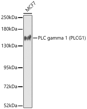
Western blot analysis of lysates from MCF7 cells using PLC gamma 1 (PLCG1) Rabbit pAb(CAB15704) at 1:1000 dilution.Secondary antibody: HRP Goat Anti-Rabbit IgG (H+L) (CABS014) at 1:10000 dilution.Lysates/proteins: 25 μg per lane.Blocking buffer: 3% nonfat dry milk in TBST.Detection: ECL Basic Kit (AbGn00020).Exposure time: 90s.