The Phospho-RelA (S536) Polyclonal Antibody (CABP0475) is a valuable tool for researchers studying NF-κB signaling pathways and gene regulation. This antibody, raised in rabbits, specifically targets the phosphorylated form of RelA at serine 536, a key post-translational modification involved in the activation of NF-κB transcription factor activity.RelA, also known as p65, is a critical subunit of the NF-κB complex, which plays a central role in regulating immune responses, inflammation, cell survival, and proliferation. Phosphorylation of RelA at S536 is a key regulatory event that promotes its translocation into the nucleus and enhances its transcriptional activity in response to various stimuli, including pro-inflammatory cytokines and oxidative stress.
The Phospho-RelA (S536) Polyclonal Antibody is validated for use in Western blot applications and is highly reactive with human, mouse, and rat samples. By specifically detecting phosphorylated RelA at S536, this antibody enables researchers to investigate the dynamics of NF-κB activation and gene expression in various cell types and physiological conditions. Its utility in immunology, cancer research, and inflammatory disorders makes it a valuable tool for studying the intricate regulation of NF-κB signaling and its implications for human health and disease.
NF-kappa-B is a ubiquitous transcription factor involved in several biological processes. It is held in the cytoplasm in an inactive state by specific inhibitors. Upon degradation of the inhibitor, NF-kappa-B moves to the nucleus and activates transcription of specific genes. NF-kappa-B is composed of NFKB1 or NFKB2 bound to either REL, RELA, or RELB. The most abundant form of NF-kappa-B is NFKB1 complexed with the product of this gene, RELA. Four transcript variants encoding different isoforms have been found for this gene.
Purification Method:
Affinity purification
Gene ID:
5970
Storage Buffer:
Store at -20℃. Avoid freeze / thaw cycles.Buffer: PBS with 0.05% proclin300,50% glycerol,pH7.3.
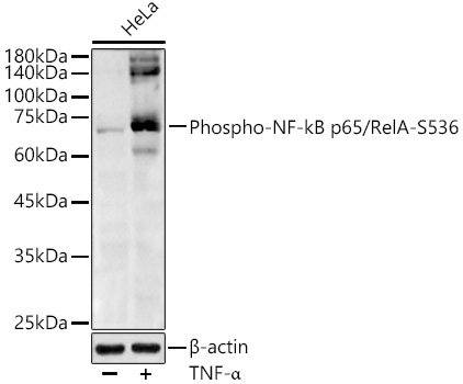
Western blot analysis of lysates from HeLa cells using Phospho-NF-kB p65/RelA-S536 Rabbit pAb (CABP0475) at 1:1000 dilution. HeLa cells were treated by TNF-α (20 ng/mL) at 37℃ for 30 minutes.Secondary antibody: HRP Goat Anti-Rabbit IgG (H+L) (CABS014) at 1:10000 dilution.Lysates/proteins: 25 μg per lane.Blocking buffer: 3% nonfat dry milk in TBST.Detection: ECL Basic Kit (AbGn00020).Exposure time: 60s.