The Phospho-NF-kB p65/RelA-S536 Antibody (CABP0124) is a high-quality antibody developed for reliable detection and analysis of target proteins. This antibody, produced in rabbits, specifically targets the phosphorylated form of RelA at serine 536, allowing for precise detection and analysis in Western blot applications.RelA, also known as p65, is a key component of the NF-κB complex and plays a central role in the activation of target genes involved in immune and inflammatory responses. Phosphorylation of RelA at serine 536 is a critical regulatory event that enhances its transcriptional activity, making it a key target for studying NF-κB signaling dynamics.
This antibody is validated for use in WB, IF/ICC, ELISA applications and has demonstrated reactivity against Human, Mouse, Rat samples.
Product Name:
Phospho-NF-kB p65/RelA-S536 Antibody
SKU:
CABP0124
Size:
20μL, 100μL
Reactivity:
Human, Mouse, Rat
Conjugate:
Unconjugated
Immunogen:
Synthetic peptide. This information is considered to be commercially sensitive.
Sequence:
FSSI A
Tested Applications:
WBIF/ICCELISA
Recommended Dilution:
WB
1:2000 - 1:10000
IF/ICC
1:50 - 1:200
ELISA
Recommended starting concentration is 1 μg/mL. Please optimize the concentration based on your specific assay requirements.
HeLa treated with TNF-α and Calyculin A, NIH/3T3 treated with TNF-α, C6 treated with TNF-α, NIH/3T3, C6
Cellular Localization:
Cytoplasm, Nucleus.
Calculated MW:
60kDa
Observed MW:
65kDa
NF-kappa-B is a ubiquitous transcription factor involved in several biological processes. It is held in the cytoplasm in an inactive state by specific inhibitors. Upon degradation of the inhibitor, NF-kappa-B moves to the nucleus and activates transcription of specific genes. NF-kappa-B is composed of NFKB1 or NFKB2 bound to either REL, RELA, or RELB. The most abundant form of NF-kappa-B is NFKB1 complexed with the product of this gene, RELA. Four transcript variants encoding different isoforms have been found for this gene.
Purification Method
Affinity purification
Gene ID
5970
RRID
AB_2771510
Buffer Information
Store at -20℃. Avoid freeze / thaw cycles. Buffer: PBS containing 50% glycerol, preserved with proclin300 or sodium azide, pH 7.3.
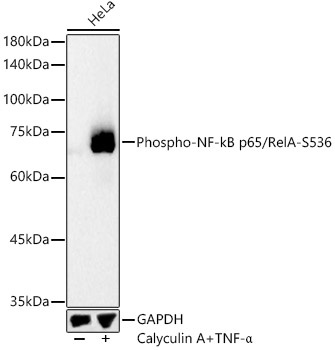
Western blot analysis of lysates from HeLa cells using Phospho-NF-kB p65/RelA-S536 Rabbit pAb (CABP0124) at 1:10000 dilution. HeLa cells were treated with TNF-α (50 ng/ml) and Calyculin A (50 nM) at 37℃ for 10 minutes. Secondary antibody: HRP-conjugated Goat anti-Rabbit IgG (H+L) (CABS014) at 1:10000 dilution. Lysates/proteins: 25 μg per lane. Blocking buffer: 3% nonfat dry milk in TBST. Detection: ECL Basic Kit (AbGn00020). Exposure time: 30s.
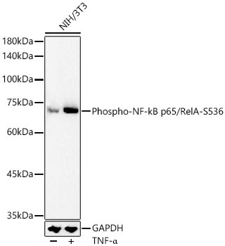
Western blot analysis of lysates from NIH/3T3 cells using Phospho-NF-kB p65/RelA-S536 Rabbit pAb (CABP0124) at 1:10000 dilution. NIH/3T3 cells were treated with TNF-α (10 ng/ml) at 37℃ for 30 minutes. Secondary antibody: HRP-conjugated Goat anti-Rabbit IgG (H+L) (CABS014) at 1:10000 dilution. Lysates/proteins: 25 μg per lane. Blocking buffer: 3% nonfat dry milk in TBST. Detection: ECL Basic Kit (AbGn00020). Exposure time: 90s.
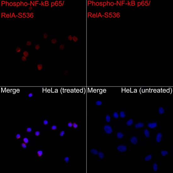
Immunofluorescence analysis of HeLa TNFα + CA and HeLa cells using Phospho-NF-kB p65/RelA-S536 Rabbit pAb (CABP0124) at dilution of 1:50 (40x lens). Secondary antibody: Cy3-conjugated Goat anti-Rabbit IgG (H+L) (CABS007) at 1:500 dilution. Blue: DAPI for nuclear staining.