The RB1 (S780) Polyclonal Antibody (CABP0444) is a valuable tool for researchers studying the phosphorylation of the RB1 protein, a key regulator of the cell cycle. This antibody, raised in rabbits, exhibits high sensitivity and specificity for detecting phosphorylation at serine 780 of the RB1 protein in human samples. Validated for use in Western blot applications, this antibody enables precise detection and quantification of RB1 phosphorylation in a variety of cell types.RB1, also known as Retinoblastoma protein, plays a crucial role in controlling cell proliferation and cell cycle progression. Phosphorylation of RB1 at serine 780 is a post-translational modification that is associated with the regulation of its tumor suppressor activity.
Research into the phosphorylation status of RB1 is essential for understanding its role in cancer development and progression, making this antibody an invaluable tool for studies in oncology and cell biology.By using the RB1 (S780) Polyclonal Antibody, researchers can gain insights into the functional significance of RB1 phosphorylation and its impact on cell cycle control and tumorigenesis. This antibody is a valuable addition to any laboratory conducting research on RB1 and its involvement in cancer biology.
Product Name:
Phospho-RB-S780 Rabbit Polyclonal Antibody
SKU:
CABP0444
Size:
20uL, 100uL
Isotype:
IgG
Host Species:
Rabbit
Reactivity:
Human,Mouse,Rat
Immunogen:
A synthetic phosphorylated peptide around S780 of human RB (NP_000312.2).
The protein encoded by this gene is a negative regulator of the cell cycle and was the first tumor suppressor gene found. The encoded protein also stabilizes constitutive heterochromatin to maintain the overall chromatin structure. The active, hypophosphorylated form of the protein binds transcription factor E2F1. Defects in this gene are a cause of childhood cancer retinoblastoma (RB), bladder cancer, and osteogenic sarcoma.
Purification Method:
Affinity purification
Gene ID:
5925
Storage Buffer:
Store at -20℃. Avoid freeze / thaw cycles.Buffer: PBS with 0.01% thimerosal,50% glycerol,pH7.3.
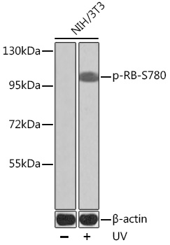
Western blot analysis of lysates from NIH/3T3 cells, using Phospho-RB-S780 Rabbit pAb (CABP0444) at dilution.NIH/3T3 cells were treated by UV at room temperature for 15-30 minutes.Secondary antibody: HRP Goat Anti-Rabbit IgG (H+L) (CABS014) at 1:10000 dilution.Lysates/proteins: 25μg per lane.Blocking buffer: 3% BSA.