The Phospho-eIF4EBP1-T37/46 Antibody (CABP0030) is a high-quality antibody developed for reliable detection and analysis of target proteins. This antibody, produced in rabbits, specifically targets the phosphorylated form of eIF4EBP1 at threonine residues 37 and 46, allowing for precise detection and analysis of this important modification.Phosphorylation of eIF4EBP1 at T37/46 is known to regulate its interaction with eukaryotic translation initiation factor 4E (eIF4E), ultimately influencing protein translation and cell proliferation. Understanding the dynamics of eIF4EBP1 phosphorylation is crucial in elucidating its role in various cellular processes, including cell cycle progression, differentiation, and survival.
This antibody is validated for use in WB, IHC-P, IF/ICC, ELISA applications and has demonstrated reactivity against Human, Mouse, Rat samples.
Product Name:
Phospho-eIF4EBP1-T37/46 Antibody
SKU:
CABP0030
Size:
20μL, 100μL
Reactivity:
Human, Mouse, Rat
Conjugate:
Unconjugated
Immunogen:
Synthetic peptide. This information is considered to be commercially sensitive.
Sequence:
STTP GGTL FSTT PG
Tested Applications:
WBIHC-PIF/ICCELISA
Recommended Dilution:
WB
1:500 - 1:1000
IHC-P
1:50 - 1:200
IF/ICC
1:50 - 1:200
ELISA
Recommended starting concentration is 1 μg/mL. Please optimize the concentration based on your specific assay requirements.
HeLa treated with IGF-1, NIH/3T3 treated with PDGF, C6 treated with PDGF
Cellular Localization:
Cytoplasm, Cytosol.
Calculated MW:
13kDa
Observed MW:
15kDa-20kDa
This gene encodes one member of a family of translation repressor proteins. The protein directly interacts with eukaryotic translation initiation factor 4E (eIF4E), which is a limiting component of the multisubunit complex that recruits 40S ribosomal subunits to the 5' end of mRNAs. Interaction of this protein with eIF4E inhibits complex assembly and represses translation. This protein is phosphorylated in response to various signals including UV irradiation and insulin signaling, resulting in its dissociation from eIF4E and activation of mRNA translation.
Purification Method
Affinity purification
Gene ID
1978
RRID
AB_2771074
Buffer Information
Store at -20℃. Avoid freeze / thaw cycles. Buffer: PBS containing 50% glycerol, preserved with proclin300 or sodium azide, pH 7.3.
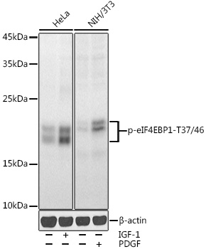
Western blot analysis of various lysates using Phospho-eIF4EBP1-T37/46 Rabbit pAb (CABP0030) at 1:1000 dilution. HeLa cells were treated with IGF-1 (50 ng/ml) at 37℃ for 30 minutes after serum-starvation overnight. NIH/3T3 cells were treated with PDGF (100 ng/ml) at 37℃ for 30 minutes after serum-starvation overnight. Secondary antibody: HRP-conjugated Goat anti-Rabbit IgG (H+L) (CABS014) at 1:10000 dilution. Lysates/proteins: 25μg per lane. Blocking buffer: 3% nonfat dry milk in TBST. Detection: ECL Enhanced Kit (). Exposure time: 30s.
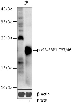
Western blot analysis of lysates from C6 cells, using Phospho-eIF4EBP1-T37/46 Rabbit pAb (CABP0030) at 1:1000 dilution. C6 cells were treated with PDGF (100 ng/ml) at 37℃ for 30 minutes after serum-starvation overnight. Secondary antibody: HRP-conjugated Goat anti-Rabbit IgG (H+L) (CABS014) at 1:10000 dilution. Lysates/proteins: 25μg per lane. Blocking buffer: 3% nonfat dry milk in TBST. Detection: ECL Enhanced Kit (AbGn00021). Exposure time: 180s.
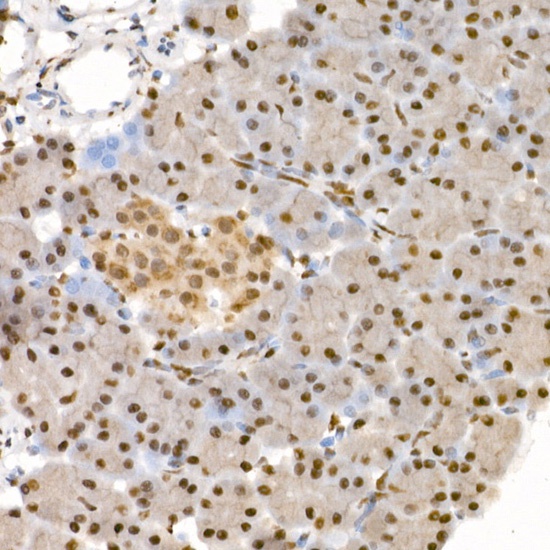
Immunohistochemistry analysis of paraffin-embedded Rat pancreas using Phospho-eIF4EBP1-T37/46 Rabbit pAb (CABP0030) at dilution of 1:100 (40x lens). High pressure antigen retrieval performed with 0.01M Citrate buffer (pH 6.0) prior to IHC staining.
Immunofluorescence analysis of U-2 OS cells using Phospho-eIF4EBP1-T37/46 Rabbit pAb(CABP0030) at a dilution of 1:100 (40x lens). Secondary antibody:Cy3 Goat Anti-Rabbit IgG (H+L)(CABS007) at 1:500 dilution. Blue: DAPI for nuclear staining.